мДЬ л°†
к∞СмГБмД† мЬ†лСРмХФ(papillary thyroid carcinoma, PTC)мЭШ нХЬ мХДнШХ м§С cribriform-morular variant(CMV)лКФ лІ§мЪ∞ лУЬлђЄ м°∞мІБнХЩм†Б мХДнШХмЬЉл°Ь, м°∞мІБк≤АмВђмГБ мВђмГБм≤і л∞П л∞©мґФм≤і л™®мЦСмЭШ мДЄнПђлУ§мЭі кіАм∞∞лРЬлЛ§[1,2]. CMV-PTCмЭШ мЮДмГБ мЦСмГБмЭА мХДмІБ лЪЬл†ЈнХШк≤М л≥ік≥†лРШлКФ к≤ГмЭА мЧЖмЬЉлВШ, 50% мЭімГБмЧРмДЬ к∞Ам°±мД± мД†мҐЕмД± мЪ©мҐЕм¶Э(familial adenomatous polyposis, FAP)мЭі лПЩл∞ШлРЬлЛ§[3].
FAPлКФ лМАмЮ•мХФмЭШ м†ДмХФмД± л≥Сл≥АмЬЉл°Ь adenomatous polyposis coli(APC) мЬ†м†ДмЮРмЭШ 5q21 л≥АмЭімЩА мЧ∞кіАлРШмЦі мЮИмЬЉл©∞, APC мЬ†м†ДмЮР л≥АнШХмЭА мЦіл¶∞ лВШмЭімЧРмДЬ лМАмЮ•мХФ л∞Ьл≥С мЬДнЧШмД±мЭД лЖТмЭілѓАл°Ь, CMV-PTC нЩШмЮРлУ§мЭА лМАмЮ• лВімЛЬк≤љк≤АмВђл•Љ нЖµнХЬ лМАмЮ• л∞П мЖМмЮ•мЭШ мЪ©мҐЕ мЧђлґА л∞П APC мЬ†м†ДмЮРмЭШ л≥АмЭі мЧђлґА к≤АмВђк∞А нХДмЪФнХШлЛ§[4]. к∞СмГБмД†мХФмЭі FAPл•Љ к∞АмІД нЩШмЮРмЭШ м≤Ђ м¶ЭмГБмЬЉл°Ь л∞ЬнШДлР† мИШ мЮИмЬЉл©∞, 4~12мДЄ мЦіл¶∞ нЩШмЮРлУ§мЭШ мХљ 30%мЧРмДЬлКФ лМАмЮ•мЪ©мҐЕмЭШ л∞ЬнШДмЭі мЮИкЄ∞ м†ДмЧР к∞СмГБмД†мХФмЭі л®Љм†А л∞Ьк≤ђлРШкЄ∞лПД нХЬлЛ§[5].
м†АмЮРлУ§мЭА к∞СмГБмД†мЭШ лЛ§л∞ЬмД± к≤∞м†ИмЭД м£ЉмЖМл°Ь лВімЫРнХЬ 16мДЄ нЩШмЮРмЭШ CMV-PTCмЩА APC мЬ†м†ДмЮР лПМмЧ∞л≥АмЭіл•Љ л≥імЭілКФ FAPк∞А лПЩл∞ШлРЬ м¶Эл°АмЧРмДЬ, мЭім†ДмЧР л≥ік≥†лРШмІА мХКмЭА APC мЬ†м†ДмЮР лПМмЧ∞л≥АмЭік∞А кіАм∞∞лРШмЦі л≥ік≥†нХШк≥†мЮР нХЬлЛ§.
м¶Э л°А
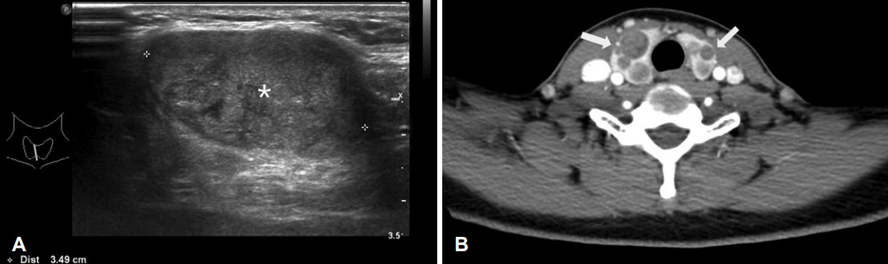
16мДЄ мЧђмЮР нЩШмЮРлКФ 2016лЕД 5мЫФмЧР м†Дк≤љлґАмЧР лІМм†ЄмІАлКФ мҐЕлђЉл°Ь лВімЫРнХШмЧђ, мЛЬнЦЙнХЬ к∞СмГБмД† міИмЭМнММ л∞П мїінУ®нД∞ м†ДмВ∞нЩФлЛ®мЄµміђмШБмГБ к∞СмГБмД† нШСлґАмЭШ 3.5 cm нБђкЄ∞мЭШ мҐЕмЦСк≥Љ мЦСмЄ° к∞СмГБмД†мЧР лЛ§мЦСнХЬ нБђкЄ∞мЭШ к≤∞м†ИмЭі мЧђлЯђ к∞Ь кіАм∞∞лРШмЧИлЛ§(Fig. 1). мЦСм†ДмЮР л∞©мґЬ мїінУ®нД∞лЛ®мЄµміђмШБ(positron emission tomography-computed tomography) мЧРмДЬлКФ к∞СмГБмД†мЧР лЛ§л∞ЬмД± мД≠мЈ® м¶Эк∞Ак∞А л≥імШАк≥†, к≤љлґАл¶ЉнФДм†ИмЭілВШ м†ДмЛ† м†ДмЭілКФ кіАм∞∞лРШмІА мХКмХШлЛ§(Fig. 2).
нЭ°мЮЕм≤ЬмЮРмДЄнПђк≤АмВђ к≤∞к≥ЉмГБ мВђмГБм≤і л∞П мГБмЛ§л∞∞ л™®мЦСмЭШ м°∞мІБмЭі кіАм∞∞лРШмЧИк≥†, л©імЧ≠мЧЉмГЙмГБ ќ≤-catenin мЦСмД± мЖМк≤ђмЭШ к∞СмГБмД†мХФмЬЉл°Ь мІДлЛ®лРШмЦі, мЭімЧР м†ДмЛ†лІИмЈ®нХШ к∞СмГБмД† м†Д м†Им†ЬмИ†мЭД мЛЬнЦЙнХШмШАлЛ§. мЬ°мХИм†БмЬЉл°Ь к∞СмГБмД† нФЉлІЙ мЩЄ мє®л≤ФмЭА л≥імЭімІА мХКмХШлЛ§.
мµЬмҐЕ л≥Сл¶ђм°∞мІБк≤АмВђ к≤∞к≥ЉмГБ, к∞СмГБмД† нШСлґАмЧР мµЬлМА 3.5√Ч3√Ч2.5 cmмЭШ мҐЕмЦСк≥Љ нХ®кїШ міЭ 18к∞Ь мЭімГБмЭШ лЛ§мЦСнХЬ нБђкЄ∞мЭШ мҐЕмЦСмЭі к∞СмГБмД† мЬ†лСРмХФмЬЉл°Ь мІДлЛ®лРШмЧИк≥†, нШДлѓЄк≤љм†БмЬЉл°Ь к∞СмГБмД† нФЉлІЙ мЩЄ мє®л≤Фк≥Љ нШИкіА мє®л≤Ф мЖМк≤ђмЭА кіАм∞∞лРШмІА мХКмХШлЛ§. мµЬмҐЕ м°∞мІБк≤АмВђ к≤∞к≥ЉлКФ мЭім†Д мДЄмє®нЭ°мЭЄк≤АмВђ к≤∞к≥ЉмЩА лІИм∞ђк∞АмІАл°Ь мГБмЛ§л∞∞ л™®мЦС(morular morphology) л∞П мВђмГБм≤і л™®мЦС(cribriform morphology)мЭШ м°∞мІБк≥Љ нХ®кїШ ќ≤-catenin л©імЧ≠мЧЉмГЙ мЦСмД±мЬЉл°Ь CMV-PTCмЧР нХ©лЛєнХЬ мЖМк≤ђмЭі л≥ік≥†лРШмЧИлЛ§(Fig. 3). мИШмИ† 2к∞ЬмЫФ мЭінЫД л∞©мВђмД† мЪФмШ§лУЬ мєШл£Мл•Љ 50 mCiл•Љ мґФк∞Ал°Ь 1м∞®л°А мЛЬнЦЙнХШмШАлЛ§.
CMV-PTCлКФ к∞Ам°±мД± мД†мҐЕмД± мЪ©мҐЕм¶ЭмЭі лПЩл∞ШлРШлКФ к≤љмЪ∞к∞А лІОкЄ∞ лХМлђЄмЧР, л≥С놕 м≤≠мЈ® л∞П мЬ†м†ДмЮР лґДмДЭмЭД мЛЬнЦЙнХШмШАлЛ§. нЩШмЮРмЭШ мХДл≤ДмІАк∞А лМАмЮ•мЭШ мЪ©мҐЕм¶ЭмЭі мЮИмЧИк≥†, лМАмЮ•мХФмЬЉл°Ь 10лЕД м†Д мВђлІЭнХЬ к∞А찱놕мЭі мЮИмЧИлЛ§. мЭімЧР мЬД л∞П лМАмЮ• лВімЛЬк≤љк≤АмВђмЩА APC мЬ†м†ДмЮР лґДмДЭмЭД мЛЬнЦЙнХШмШАлЛ§. мЬД лВімЛЬк≤љк≤АмВђмГБ, мЬДм†А мЪ©мҐЕ(stomach fundic polyp) л∞П лМАмЮ• лВімЛЬк≤љк≤АмВђмГБ мЖМмЮ•мЧРмДЬ 1к∞Ь, лМАмЮ•мЧРмДЬ 15к∞ЬмЭШ мД†мҐЕмЭі кіАм∞∞лРШмЧИлЛ§. APC мЬ†м†ДмЮРмЧР лМАнХЬ мЧЉкЄ∞мДЬмЧі лґДмДЭ к≤∞к≥Љ, 5л≤И мЧЉмГЙм≤і q armмЭШ 21л≤И мЬ†м†ДмЮР 4181~4188л≤ИмІЄ мЧЉкЄ∞мДЬмЧімЭі к≤∞мЖР(deletion)лРШмЦі 1394л≤ИмІЄ мХДлѓЄлЕЄмВ∞мЭЄ мХДмК§нММнКЄмВ∞(Aspartic acid)мЭі кЄАл¶ђмЛ†(Glycine)мЬЉл°Ь мєШнЩШлРШл©імДЬ, м°∞кЄ∞ мҐЕк≤∞(premature termination)лРШлКФ нФДл†ИмЮДмЛЬнФДнКЄ лПМмЧ∞л≥АмЭі(frameshift mutation)к∞А кіАм∞∞лРШмЧИлЛ§(Table 1, Fig. 4). мЭі лПМмЧ∞л≥АмЭілКФ мЭім†ДмЧР л≥ік≥†лРЬ м†Б мЧЖлКФ мГИл°ЬмЪі мДЬмЧі л≥АнЩФмЭілЛ§[6].
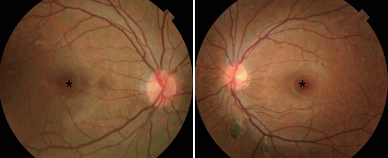
нЩШмХДлКФ лЛ§л∞ЬмД± к∞СмГБмД†мХФ л∞П к∞Ам°±мД± мД†мҐЕмД± мЪ©мҐЕм¶Эк≥Љ лНФлґИмЦі мЛЬ놕 м†АнХШл•Љ нШЄмЖМнХШмЧђ мЛЬнЦЙнХЬ мХИк≥Љ к≤АмІДмГБ мҐМмХИ лІЭлІЙ нХШл∞©мЬЉл°Ь мГЙмЖМк∞А мє®м∞©лРЬ мД†м≤ЬмД± лІЭлІЙ мГЙмЖМ мє®м∞©мД± мГБнФЉ лєДлМАм¶Э(congenital hypertrophy of the retinal pigment epithelium, CHRPE) мЖМк≤ђмЭі кіАм∞∞лРШмЧИк≥†, кЈЄ мЩЄ лЛ§л•Є нФЉлґА л≥Сл≥АмЭА кіАм∞∞лРШмІА мХКмХШлЛ§(Fig. 5).
мИШмИ† нЫД 1лЕДмІЄ, нЩШмЮРлКФ міИмЭМнММк≤АмВђ л∞П лПЩмЬДмЫРмЖМк≤АмВђ лУ±мЭД нЖµнХі м†ХкЄ∞к≤АмІДмЭД мЛЬнЦЙнХШк≥† мЮИмЬЉл©∞, мЮђл∞ЬмЭА кіАм∞∞лРШмІА мХКмХШлЛ§. лМАмЮ• мД†мҐЕмЧР лМАнХімДЬлКФ к≥†лУ±нХЩкµР м°ЄмЧЕ мЭінЫДмЧР мИШмИ†м†Б мєШл£Мл•Љ к≥†л†§нХШк≥† мИШмИ† м†ДкєМмІА 1лЕДлІИлЛ§ лМАмЮ• лВімЛЬк≤љ л∞П мЬД мЛЭлПД лВімЛЬк≤љмЭД мЛЬнЦЙнХШк≥†, мҐМмЄ° лИИ л≥Сл≥АмЧР лМАнХімДЬлПД 1лЕДлІИлЛ§ м†ХкЄ∞к≤АмІДмЭД мЛЬнЦЙнХ† мШИм†ХмЭілЛ§.
к≥† м∞∞
CMV-PTCлКФ к∞СмГБмД† мЬ†лСРмХФмЭШ мХДнШХмЬЉл°Ь лґДл•ШлРШмІАлІМ, м†ДнШХм†БмЭЄ мЬ†лСРмХФк≥Љ кµђл≥ДлРШлКФ нКємІХм†БмЭЄ мЮДмГБ мЦСмГБмЭД к∞АмІАк≥† мЮИлЛ§. к∞СмГБмД† мЬ†лСРмХФмЭА мµЬкЈЉ World Health Organization лґДл•Шл≤ХмЧР мЭШнХШмЧђ 15к∞АмІАл°Ь лґДл•ШлРШл©∞, мЭі м§С CMV-PTCлКФ 0.2%л•Љ м∞®мІАнХШлКФ лУЬлђЄ мХДнШХмЭілЛ§[7].
к∞СмГБмД† мЬ†лСРмХФмЭі 40лМАмЧР нШЄл∞ЬнХШлКФ л∞Шл©і CMV-PTCлКФ 20лМАмЧР нШЄл∞ЬнХШл©∞[3], л≥Є м¶Эл°АмЩА к∞ЩмЭі 10лМАмЧРлПД лєДкµРм†Б нШЄл∞ЬнХШлКФ к≤ГмЬЉл°Ь мХМ놧솪 мЮИлЛ§. лШРнХЬ к∞СмГБмД† мЬ†лСРмХФмЭА л¶ЉнФДм†И м†ДмЭімЭШ мЬ†л≥С땆мЭі лЖТмЭА л∞Шл©імЧР, CMV-PTCлКФ мХљ 10% м†ХлПДмЭШ л¶ЉнФДм†И м†ДмЭіл•Љ л≥імЭіл©∞ мЫРк≤© м†ДмЭімЭШ мЬ†л≥С땆лПД лВЃк≤М кіАм∞∞лРШмЧИлЛ§[3,8].
CMV-PTCк∞А мЮДмГБм†БмЬЉл°Ь к∞АмЮ• нКємІХм†БмЭЄ к≤ГмЭА FAPмЩАмЭШ мЧ∞кіАмД±мЭілЛ§. FAPлКФ мГБмЧЉмГЙм≤і мЪ∞мД± мІИнЩШмЬЉл°Ь, лМАмЮ•мЭШ лЛ§л∞ЬмД± мЪ©мҐЕк≥Љ APC мЬ†м†ДмЮРмЭШ 5л≤И мЧЉмГЙм≤і q armмЭШ 22л≤И мЬ†м†ДмЮРмЧРмДЬ л≥АмЭік∞А кіАм∞∞лРЬлЛ§. FAP нЩШмЮРлКФ к±∞мЭШ лМАлґАлґДмЭі лМАмЮ•мХФмЬЉл°Ь мІДнЦЙнХШл©∞, мЦіл¶∞ лВШмЭімЧР лЛ§л∞ЬмД±мЬЉл°Ь л∞ЬмГЭнХ† мИШ мЮИлЛ§. кЄ∞м°імЭШ л©ФнГАлґДмДЭмЧРмДЬлКФ 53%мЭШ CMV-PTC нЩШмЮРмЧРмДЬ FAPк∞А лПЩл∞ШлРШмЧИмЬЉл©∞, мЭі м§С мХљ 50%мЧРмДЬ к∞СмГБмД†мХФмЭі л®Љм†А мІДлЛ®лРШмЧИлЛ§[3,9]. л≥Є нЩШмЮРмЭШ к≤љмЪ∞ APC gene mutation нЩХмЭЄмЭД мЬДнХШмЧђ, лІРміИнШИмХ°мЭШ л∞±нШИкµђл°ЬлґАнД∞ Genomic DNAл•Љ мґФмґЬнХШмЧђ 5л≤И мЧЉмГЙм≤імЭШ q armмЭШ 21~22л≤ИмІЄмЧР мЬДмєШнХЬ APC мЬ†м†ДмЮРмЭШ л™®лУ† coding exon л∞П мЭЄм†С intron лґАмЬДмЭШ мЧЉкЄ∞мДЬмЧімЭД direct sequencingмЭД нЖµнХЬ мЧЉкЄ∞мДЬмЧі лґДмДЭл≤ХмЬЉл°Ь нЩХмЭЄнХШмШАлЛ§. міЭ 15к∞Ь exonмЭШ мЧЉмГЙм≤і лґДмДЭмЧРмДЬ APC мЬ†м†ДмЮРмЭШ 5л≤И мЧЉмГЙм≤і q armмЭШ 21л≤И мЬ†м†ДмЮР 4181~4188л≤ИмІЄ мЧЉкЄ∞мДЬмЧімЭі deletion лРШл©імДЬ Aspartic acidк∞А GlycineмЬЉл°Ь мєШнЩШлРШлКФ л≥АмЭік∞А кіАм∞∞лРШл©імДЬ, APC мЬ†м†ДмЮРк∞А м°∞кЄ∞мЧР мҐЕк≤∞лРШмЧИлЛ§. лМАлґАлґД л∞ЭнШАмІД APC gene mutationмЭШ к≤љмЪ∞, 3927~3931 мЧЉкЄ∞мДЬмЧімЭі deletion лРШл©імДЬ frameshift mutationмЭі л∞ЬмГЭнХШмЧђ APC мЬ†м†ДмЮРмЭШ premature terminationмЧР мЭШнХі л≥АмЭік∞А лРЬлЛ§к≥† л∞ЭнШАм†Є мЮИмЬЉлВШ, л≥Є м¶Эл°АмЧРмДЬлКФ кЄ∞м°ік≥ЉлКФ лЛђл¶ђ 4181~4188л≤И мЧЉкЄ∞мДЬмЧімЭШ deletionмЧР мЭШнХЬ APC мЬ†м†ДмЮР лПМмЧ∞л≥АмЭіл°Ь, кЄ∞м°імЧР л≥ік≥†лРШмІА мХКмЭА мЬ†м†ДмЮР л≥АмЭімЭілЛ§.
мЭімЩА лНФлґИмЦі FAPл•Љ к∞АмІД нЩШмЮРмЭШ к≤љмЪ∞ мХљ 75%мЧРмДЬ CHRPEк∞А лПЩл∞ШлРШк≥† мЮИмЬЉл©∞, нКєнЮИ мЦСмЄ° лІЭлІЙмЭШ мГЙмЖМ мє®м∞© нШємЭА л≥Сл≥АмЭШ м†АмГЙмЖМм¶ЭмЭі лПЩл∞ШлРЬ к≤љмЪ∞мЧРлКФ FAPмЩА мЧ∞кіАлРЬ CHRPEмЭШ мІАнСЬлЭЉк≥† нХ† мИШ мЮИмЦі CMV-PTC нЩШмЮРмЭШ к≤љмЪ∞, м†ХкЄ∞м†БмЬЉл°Ь лІЭлІЙк≤АмВђк∞А нХДмЪФнХШлЛ§[10].
CMV-PTCлКФ м°∞мІБнХЩм†БмЬЉл°Ь cribriformмЭШ кµђм°∞л•Љ к∞АмІАк≥† мЮИк≥†, morule нШХнГЬмЭШ мДЄнПђ кµђм°∞л•Љ к∞АмІАк≥† мЮИмЬЉл©∞[1,2], ќ≤-catenin л©імЧ≠мЧЉмГЙмЧРмДЬ нХµк≥Љ мДЄнПђмІИмЭі мЧЉмГЙлРШлКФ к≤ГмЭД нЩХмЭЄнХ®мЬЉл°ЬмН® мІДлЛ®мЭД нХ† мИШ мЮИлЛ§[11].
CMV-PTCмЭШ мєШл£Мл≤ХмЭА мЬ†л≥С땆мЭі лВЃмХД м†Хл¶љлРШмІАлКФ мХКмХШмІАлІМ, мЧђнГА к∞СмГБмД† мЬ†лСРмХФмЭШ мєШл£М мЫРмєЩмЭД лФ∞л•ЄлЛ§. л≥Є м¶Эл°АмЩА к∞ЩмЭі лЛ§л∞ЬмД± к∞СмГБмД†мХФмЭі нЭФнХШкЄ∞ лХМлђЄмЧР к∞СмГБмД† м†Д м†Им†ЬмИ†мЭД мЛЬнЦЙнХШл©∞, л¶ЉнФДм†И м†ДмЭік∞А нЩХмЭЄмЭі лРШл©і л¶ЉнФДм†И м†Им†ЬмИ†мЭД мґФк∞АнХШк≤М лРЬлЛ§[3,9,12]. л≥Є м¶Эл°АмЧРмДЬлКФ к∞СмГБмД†мЭШ лЛ§л∞ЬмД± мҐЕмЦСмЧР лМАнХі м†Д к∞СмГБмД† м†Им†ЬмИ†мЭД мЛЬнЦЙнХШмШАк≥†, лМАмЮ• мЪ©мҐЕмЧР лМАнХімДЬлКФ к≤љк≥ЉкіАм∞∞ нЫД FAPмЭШ мєШл£М мЫРмєЩмЧР лФ∞лЭЉ нЩШмХД мД±мЮ• мЭінЫД лМАмЮ•мЭШ лґАлґД м†Им†ЬмИ†мЭД к≥†л†§нХШк≥† мЮИлЛ§.
л≥Є м¶Эл°АмЧРмДЬлКФ лУЬлђЉк≤М л≥ік≥†лРШк≥† мЮИлКФ CMV-PTC нЩШмЮРмЧРмДЬ м†ДнШХм†БмЭЄ мЮДмГБ мЦСмГБмЭД к∞Ам°МмЬЉлВШ, мЭім†ДмЧР л≥ік≥†лРШмІА мХКмХШлНШ мГИл°ЬмЪі APC мЬ†м†ДмЮРмЭШ л≥АнШХмЭі кіАм∞∞лРШмЧИк≥†, FAPк∞А лПЩл∞ШлРЬ м¶Эл°Ал•Љ мІДлЛ® л∞П мєШл£МнХШмШАкЄ∞мЧР л≥ік≥†нХШмШАлЛ§. CMV-PTCлКФ к∞СмГБмД†мХФмЭШ нКємІХм†БмЭЄ мЮДмГБ мЦСмГБк≥Љ лНФлґИмЦі, лМАмЮ•мХФмЭШ м†ДмХФмД± л≥Сл≥АмЬЉл°Ь мХМ놧мІД FAPк∞А нЭФнХШк≤М лПЩл∞ШлРШкЄ∞ лХМлђЄмЧР мЭімЧР лМАнХЬ к∞Бл≥ДнХЬ м£ЉмЭШк∞А нХДмЪФнХШк≤†лЛ§.