Introduction
Lipoma is a benign neoplasm which grows slowly, which occurs in the neck, trunk, and extremities, but rarely found in the nasal sinus and nasal septum [1]. Until now, lipomas of the nasal septum were reported in one case in Korea [2], and three cases (including fibrolipoma, which is a subtype of lipomas) abroad [1,3,4]. We found a mass in the nasal septum who presented with intermittent nasal obstruction. The mass was completely removed by endoscopic endonasal surgery, and histological examination revealed a fibrolipoma. We report this case with a review of the literature.
Case
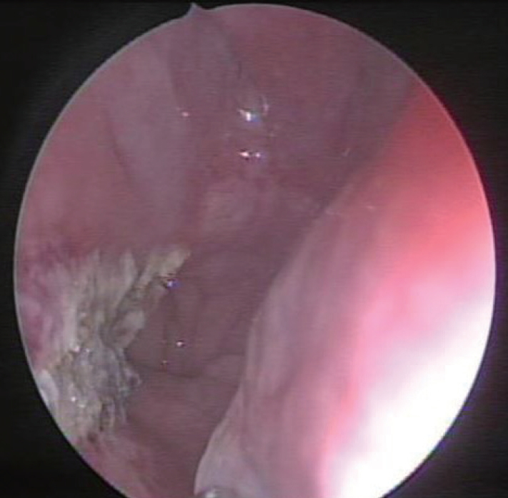
Thirty-two-year-old woman visited us with complaints of intermittent nasal obstruction and throat discomfort. She had no other symptoms nor underlying disease. An endoscopic examination revealed an elongated round mass covered by normal nasal mucosa originated from the posterior edge of the nasal septum that extended to the nasopharynx (Fig. 1A and B). Computed tomography (CT) revealed a 2.7Г—1.5 cmsized, low-density, polypoid mass originating from the posterior part of the nasal septum with an elongated neck occupying the left nasopharyngeal to oropharyngeal cavity (Fig. 1C). The nasal septum slightly deviated to the right side and concha bullosa in the left middle turbinate was observed (Fig. 1D).
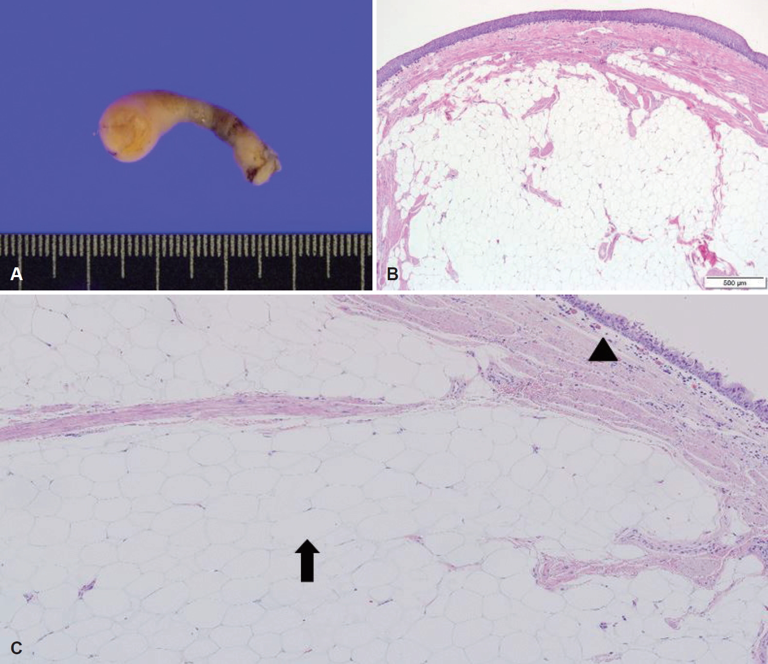
We planned to conduct an endoscopic endonasal mass excision surgery. After the induction of general anesthesia, 1:1000 epinephrine mixed with lidocaine was injected into both septal mucosa. After correcting the deviated nasal septum, we located the mass in the nasopharynx which measured approximately 3.0Г—1.0 cm. We completely removed the mass and a silastic sheet was placed on both of the nasal septum and fixed using vicryl 4-0. On histopathological examination, the mass was approximately 2.7Г—1.5 cm and consisted of homogeneously-distributed yellow tissue which had a clear margin with the surrounding tissue (Fig. 2A). Microscopically, mature adipose tissue cells were found in the subepithelium; no atypical cells were found (Fig. 2B), and the surface of the mass was covered with ciliated pseudostratified columnar epithelium cells which appeared to be normal respiratory epithelium (Fig. 2C). The final histological diagnosis was fibrolipoma. Postoperatively, the mucosa of the posterior nasal septum recovered to normal, and the patient was followed for more than 6 months without any recurrence (Fig. 3).
Discussion
Lipoma is a slowly growing benign tumor and consists of mature fat cells, which usually occurs in the neck, trunk, or limbs. However, lipomas originated from the parasinus and nasal septum are extremely rare. Because there is a paucity of adipose in the parasinus area, the tissues are less likely to progress to lipomas [3,4]. The lipomas remain asymptomatic clinically, but once they grow in size, which can cause discomfort as they compress surrounding structures [5]. Lipomas originated from the nasal septum result in symptoms such as unilateral nasal obstruction, facial edema, tenderness, rhinorrhea and epistaxis [2].
The reported benign tumors found in the nasal septum include schwannoma, pleomorphic adenoma, chondroma, hemangioma, teratoma, leiomyoma, papilloma, and lipoma [2]. Lipomas are classified according to their subtypes, which include simple lipomas, fibrolipomas, myxoid lipomas, angiolipomas, spindle cell lipomas, pleomorphic lipomas, angiomyolipomas, myelolipomas, and lipoblastomatosis [6,7]. The microscopic findings in this case revealed that the surface of the mass was covered with ciliated pseudostratified columnar epithelium cells which appeared to be normal respiratory epithelium, and mature adipose tissue cells were found in the subepithelium, that revealed fibrolipoma. The causes of lipomas have not been evaluated yet. It is known to be caused by the remaining adipose tissues during progress or development of existing adipocytes [8]. 1% of fibrolipomas occur in the facial region [1], and fibrolipomas that originated in the nasal septum reported in one adult [1] and one child [9] worldwide. In addition, lipomas in the parasinus can occur very rarely in infants as part of a syndrome that includes symptoms involving the brain [10]. Fibrolipomas are mostly diagnosed as benign, and progression to liposarcoma is extremely rare [1].
Preoperative radiologic examination is essential to differentiate between other tumors and lipomas and to know the exact extension of the mass [4]. In particular, CT and magnetic resonance imaging (MRI) help diagnose lipoma and discover its extension and characteristics. The radiologic characteristics of lipomas are as follows: low-density findings on CT, high signal intensity on T1-weighted MRI, and low signal intensity on T2-weighted MRI [4]. In this case, the CT images showed low-density polypoid findings.
Treatment for facial lipomas is the same as for those which occur in other parts of the body: complete surgical resection [7,9]. In this case, the mass and the posterior margin of the nasal septum were completely removed with a 1 cm resection margin. We endeavored to preserve as much of septal mucosa as possible, and the mass with capsule was well separated from the mucosa of the septum. Blood vessels which were exposed during surgery were cauterized by the bipolar cauterizer.
Lipomas can relapse several years after they have been completely removed, but local recurrence rate is less than 5%. Therefore, long-term follow-up must be recommended [11,12].
We report a case of fibrolipoma originating from the posterior part of the nasal septum with an elongated neck occupying the left nasopharyngeal to oropharyngeal cavity, which was successfully removed by an endoscopic approach, and we also report a review of the literature.