Introduction
Endoscopic sinus surgery (ESS) is a common surgical treatment for chronic sinusitis. Although the navigation system reduces the risks in an endoscopic surgery, the wide anatomical diversity limits the application of endoscopy. Therefore, it is essential to understand the developmental state and variations of the paranasal sinuses (PNSs) and the other anatomical structures. The developmental state of the frontal sinus (FS) can be classified into aplasia, hypoplasia, and control groups. There is limited literature between the relation of the degree of FS pneumatization and the development of the PNS structures [1-4].
Lateral lamella length (LLL) and olfactory fossa depth are important anatomical parameters to be considered in preventing surgical complications like skull base injury. Greater LLL increases the possibility of lateral lamella injury during the surgery. During ESS, Keros type III fossa is at a greater risk for skull base injury than type I fossa [5]. A few studies have reported a direct relationship between the pneumatization of FS and the LLL [1,2]. In contrast, another study has reported that lesser the pneumatization of the FS, greater is the LLL [3]. These results could be a matter of contention.
Thus, in this study, we focused on two points. Using computed tomography (CT) multiplanar reconstruction (MPR), first, we investigated the anatomical variants of the PNS and its association with the state of FS pneumatization. Second, we studied the correlation between the degree of pneumatization of the FS, maxillary sinus (MS), and sphenoid sinus (SS) and the LLL.
Subjects and Methods
Subjects
We analyzed 966 PNS CT scans that were conducted at our university hospital from July 2017 to December 2018. Patients aged 18 to 70 years were included. Of 802 PNS CT scans, we excluded the patients with surgical history (440 cases), trauma history (28 cases), tumor (16 cases), or an expansile lesion (7 cases). We included total 311 PNS CT scans for the study. After excluding patients with surgical history, trauma history, tumor, or an expansile lesion, 311 PNS CT scans were considered for the study. The approval of the Institutional Review Board of Wonkwang University Hospital was obtained (2020-06-011-001).
Analysis using high resolution sinus tomography
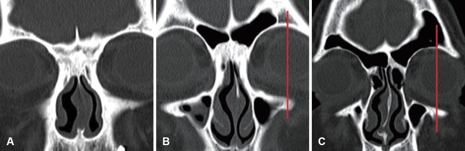
We analyzed 311 PNS CT scans (n=622, including left and right sides). Tomography was performed using Somatom Definition Flash 256-slice CT scanners (Siemens Healthcare, Forchheim, Germany) with axial plane reconstruction of 1 mm slice thickness. We classified FS into type I (aplasia), type II (hypoplasia), and type III (control) (Fig. 1) [1]. In type I, the FS does not pneumatize on the orbital roof at all. Type II shows pneumatization limited to the medial aspect of the midorbital line. FS pneumatization above the orbital roof is defined as type III. Based on the classification by Bolger, et al. [6], the MS was assigned as control and hypoplasia. Type I hypoplasia is characterized by a normal uncinate process, well-defined infundibular passage, and mild sinus hypoplasia. Type II is characterized by absence or hypoplasia of the uncinate process, an ill-defined infundibular passage, and soft-tissue density opacification of a significantly hypoplastic sinus. Type III shows absence of the uncinate process and a profoundly hypoplastic, cleft-like sinus. In our study, we did not come across type III, and thus classified type I and II as hypoplasia of the MS. The SS was classified into sellar and presellar types.
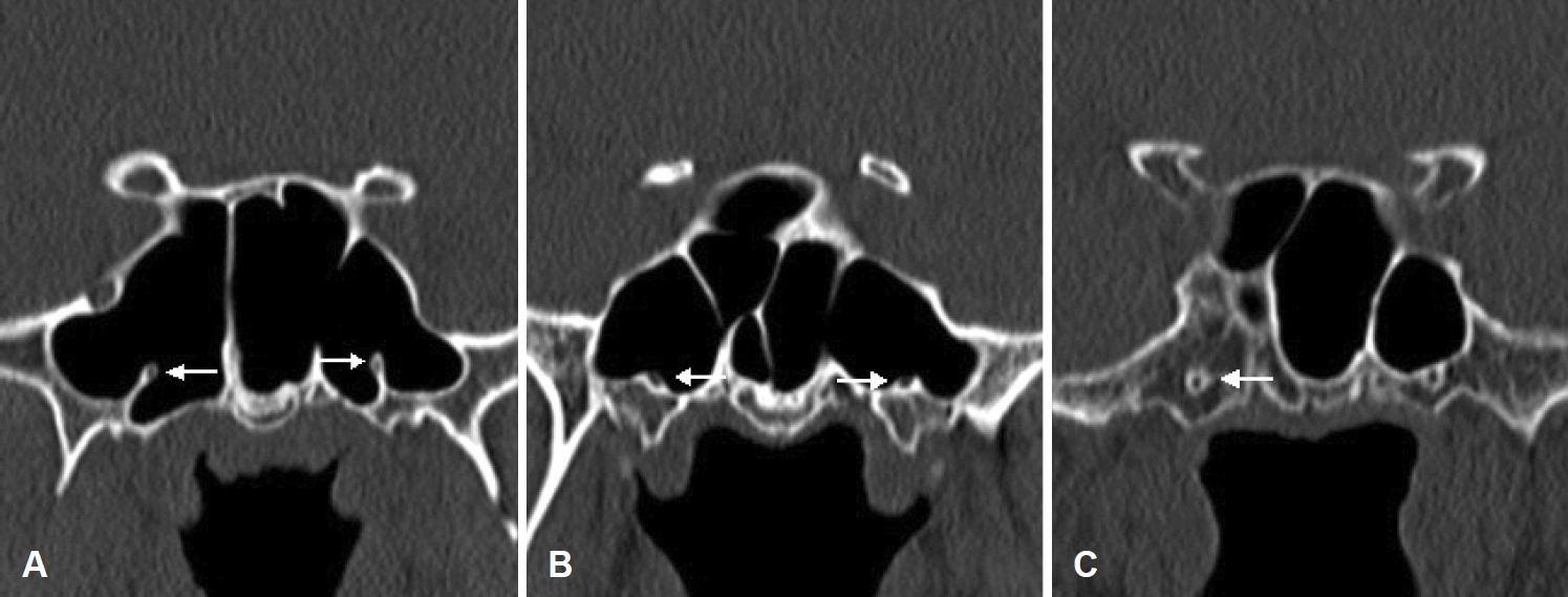
We investigated superior turbinate pneumatization (STP), middle turbinate pneumatization (MTP), agger nasi cell (ANC), infraorbital cell (IOC), optic nerve (ON) type, and vidian nerve (VN) type. We categorized ON canals into four types using the classification system by Itagi, et al. [7] (Fig. 2). Type I canal is superolateral to the SS and does not indent on the sinus wall, as seen on the coronal CT planes. Type II canal is indented on the SS contour and the indented portion is less than 50% of the nerve circumference. Type III canal traverses through the SS and coronal sections showing more than 50% projection of ON circumference into the SS. Type IV is a canal adjacent to the SS and posterior ethmoid sinuses/Onodi cell. Configurations of the VN were classified into three types (Fig. 3). Type I is considered when the VN canal completely protrudes into the SS; type II, when the canal partially protrudes into the SS or into the floor of the SS; and type III, when the canal is completely obscured within the sphenoid corpus [8].
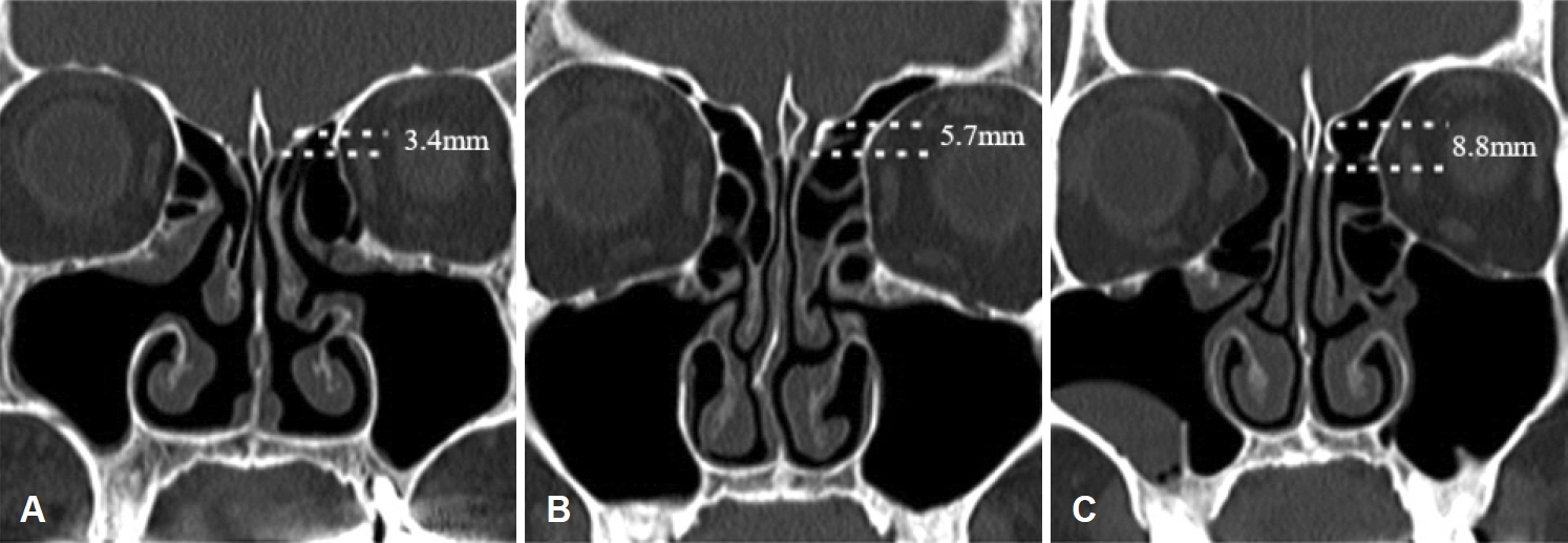
The LLL was measured as the distance between the fovea ethmoidalis and the horizontal cribriform plate in the olfactory fossa, as observed on the coronal plane in which the crista galli was well observed (Fig. 4) [3]. Two otolaryngologists analyzed each case, and on disagreement, a unified opinion was considered after referring to a third otolaryngologist.
Statistics
Statistical analyses were performed using IBM SPSS Statistics (version 24) (IBM Corp., Armonk, NY, USA). Chi-square test and FisherвАЩs exact test were applied for the evaluation of anatomical structures and variations of paranasal structures among the three groups of the FS. One-way analysis of variance method was used to compare the LLL among the three groups of the FS. Post-hoc comparisons were conducted using ScheffeвАЩs test. Student independent t-test was performed to determine the relation between the LLL and the hypoplastic and control MS and the presellar and sellar types of SS. A p value of <0.05 was considered to indicate statistical significance.
Results
Subjects
Out of 622 sides that were analyzed, 52 FS were noted as aplasia, 271 as hypoplasia, and 299 as control. The mean age of the patients was 50.88±14.39 years. The average ages of the aplasia, hypoplasia, and control groups were 57.06±9.45, 52.81±14.00, and 48.07±14.86 years, respectively. There were 169 men (54.3%) and 142 women (45.7%). Among men, FS type I, II, and III were observed in 22, 129, and 187 cases, respectively. In women, FS type I, II, and III were observed in 30, 142, and 112 cases, respectively. We observed more women with FS type I and more men with type III, and there was a significant difference seen between men and women (p<0.001) (Table 1).
STP, MTP, ANC, and IOC prevalence according to the FS type
STP was noted with 7, 97, and 107 cases among FS type I, II, and III, respectively. Both, type II and III, showed more frequent occurrences of STP than type I (p=0.005). In type I, 10 cases showed MTP, while 88 and 102 cases showed MTP in FS type II and III, respectively. There were no significant differences among all the types (p=0.105). All 52 cases of FS type I revealed ANC. In FS type II and III, there were 261 and 292 cases with ANC, respectively, with no significant difference observed (p=0.278). IOC was noted in 4, 38, and 48 cases among FS type I, II and III, respectively. There were no significant differences among them (p=0.276) (Table 1).
MS and SS pneumatizations according to the FS type
In FS type I, II, and III, MS was controlly pneumatized in 44, 236, and 275 cases, respectively. Furthermore, in FS type I, II, and III, MS hypoplasia occurred in 8, 35, and 24 cases, respectively. There were no significant differences observed (p=0.091). FS type I, II, and III showed 20, 40, and 30 cases of presellar SS while sellar SS was noted with 32, 231, and 269 cases, respectively. No significant difference was observed (p=0.057) (Table 1).
ON and VN types according to the FS type
ON type I, II, III, and IV were present in 32, 10, 1, and 9 cases, respectively, in FS type I cases; 136, 72, 13, and 50 cases, respectively, in FS type II cases; and 95, 85, 38, and 81 cases, respectively, in FS type III cases. The ON type III and IV were significantly frequent in FS type III (p<0.001). In FS type I, there were 5 cases of VN type I, 10 cases of type II, and 37 cases of type III. FS type II cases revealed 54, 95, and 122 cases of VN type I, type II, and type III, respectively. VN type I, II, and III were noted in 97, 119, and 83 cases, respectively, among FS type III. The occurrence of VN type III in FS type I was significant (p<0.001) (Table 1).
Discussion
In this study, we attempted to confirm our speculation about a possible correlation between the pneumatization of FS and the development of other PNS structures. To the best of our knowledge, there are only few related studies in the literature.
In our study, STP rate was significantly higher in type II and III than I. However, MTP rate according to FS type was not significant. In the study by Yazici [4], STP rate was 12.5% in FS type I, 30% in type II, and 50% in type III, significantly higher in type III. In their study, MTP rate was 44.2%; 30.0% in FS type I, 40.0% in type II, and 62.5% in type III, significantly higher in type III. Unlike our study, Yazici [4] showed significant differences in MTP rate; this is probably because their study targeted Turkish and our study Koreans. That is, it may have been caused by racial and geographical differences. The study by Yazici [4] showed similar results, indentation rates of ON and VN being higher in the FS type III than in type I or II. The ON types III and IV are more susceptible to damage during ESS.
Greater LLL has a significant clinical implication. The lateral lamella of the cribriform plate is the thinnest part of the skull base, known as the common injury area during ESS [9]. The thickness of the lateral lamella is known to be only 0.05 mm. Greater tendency of damage is suspected with an increase in the length of lateral lamella; hence, great caution is suggested during the operation.
In our study, the mean LLL of type II and III were significantly greater than that of type I but the MS and SS types and the LLL were not significantly correlated. Kayabasi, et al. [3] observed that the pneumatization defects of MS, FS, and SS were related to deeper olfactory fossa and longer lateral lamella. In their study, the mean LLL of the FS aplasia group was 6.53¬±1.78 mm, 5.98¬±1.94 mm in the FS hypoplasia group, and 4.28¬±1.52 mm in the control FS group (p<0.001). They found that patients with MS and SS hypoplasia had a deeper olfactory fossa and longer lateral lamella. However, a few studies showed similar results to ours [1,2]. √ЗomoƒЯlu, et al. [1] reported deeper olfactory fossa in extensively pneumatized FS cases, with a mean LLL of 4.17¬±1.44 mm in FS type I, 5.15¬±1.50 mm in type II, and 5.60¬±1.41 mm in type III. Kayabasi, et al. [3] and two other studies [1,2] surveyed the Turkish people and obtained different results.
The limitation of this study is its small number of FS type I compared to type II and III, which is a very small representation of the general population. The development of the FS may differ depending on gender, race, and region. Bilateral FS aplasia was reported to vary from 1.3 to 43.0% in previous studies. In a Korean study by Sim, et al. [10], bilateral FS aplasia was observed in 2.9% of men and 7.1% of women. Unilateral FS aplasia has been reported to vary from 0.8 to 14.3% in different studies. Sim, et al. [10] reported an incidence of 5.4 and 5.3% in men and women, respectively. This relatively low rate of FS aplasia is consistent with our results. We studied limited CT scans that were conducted from July 2017 to December 2018, thus accounting to a small sample size. A larger number of PNS CT scans need to be analyzed for further studies.
In conclusion, this study correlated the variations of PNS structures to the FS development and compared the LLL according to the FS, SS, and MS pneumatization. In our study we observed greater indentation of the ON and VN into the SS and a longer LL with increasing FS pneumatization. Therefore, surgeons should be more cautious and carefully review the CT scan to avoid damage of the important structures such as the LLL, ON, and VN, in cases of well pneumatized FS.