Introduction
Sinusitis occurs widely at any age, regardless of sex or race, and chronic sinusitis has a prevalence of 1%-12%, according to diagnostic criteria[1]. The main symptoms of sinusitis include nasal congestion, rhinorrhea, decreased sense of smell, and facial discomfort. However, sinusitis is a heterogeneous inflammatory disease that sometimes shows unique symptoms without the above-named cardinal symptoms.
In particular, rhinologic symptoms do not often appear in the case of the posterior sinus involvement, while unusual symptoms such as headache or visual symptoms appear [2]. Thus, the clinician sometimes misdiagnoses or delays treatment. The structures adjacent to the posterior sinus, such as the optic nerve, internal carotid artery, and cavernous sinus, are affected, and mortality rate of 2.9% has been reported [3].
Onodi cells, located laterally and superiorly into the sphenoid sinus, may often aerate the anterior clinoid process (ACP). Onodi cells and the aerated ACP are also in contact with important structures, and neurological symptoms due to inflammation and tumors of these structures have been reported [4]. Since sinusitis is not common as a cause of isolated cranial palsy, it is easy to overlook it unless sufficient suspicion is preceded [5].
We report the case of one patient with Onodi cell sinusitis with isolated oculomotor nerve palsy who underwent endoscopic sinus surgery and recovered completely after 2 months.
Case
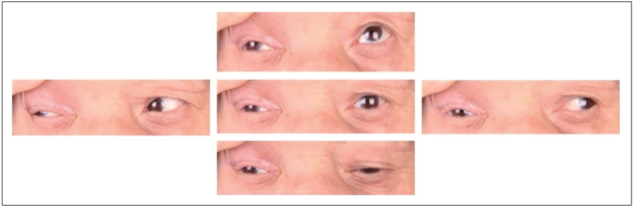
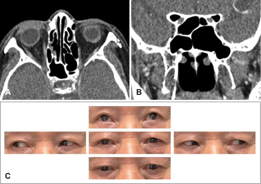
A 75-year-old male patient visited our neurology department with persistent double vision and headache for 3 days. The patient had a history of hypertension and diabetes. Physical examination revealed that the right ptosis and exotropia of the right eye. Visual acuity and intraocular pressure were normal, and no abnormalities were observed in bilateral pupil control. In the extraocular muscle examination, there was a complaint of horizontal diplopia between 45 degrees to the right and 180 degrees to the left during binocular gaze and adduction, supination, and downward restriction of the right eye, and worsening on left gaze (Fig. 1).
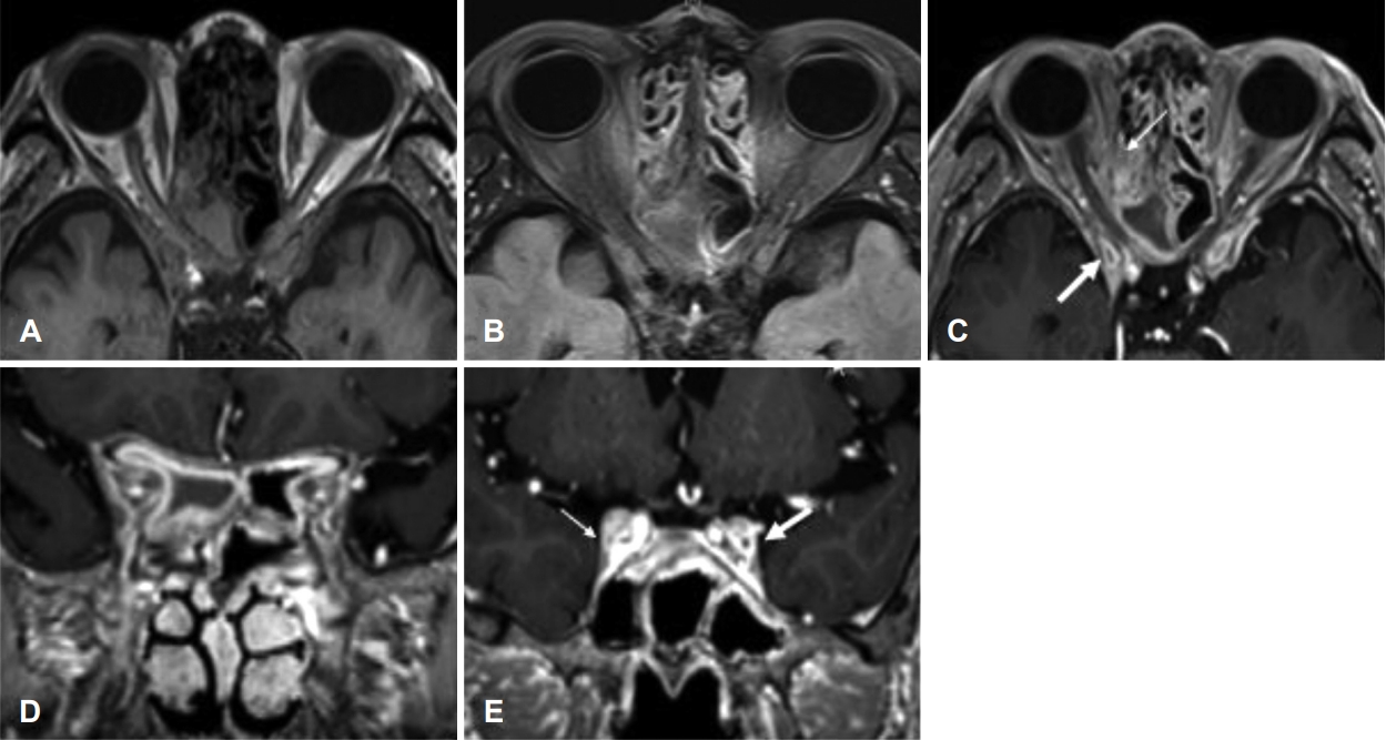
There were no significant abnormalities in laboratory test except HbA1c, slightly increased to 7.2. On the day of admission, brain MRI showed past lacunar cerebral infarction but no lesions in the brain parenchyma that could explain the symptoms were observed. On the right Onodi cell and ACP, brain MRI showed intermediate on T1, intermediate to low signal on T2, enhanced mucosa on the peripheral wall in gadolinium imaging and extraconal haziness were found (Fig. 2A-D). Compared with the normal left side, the right oculomotor nerve was compressed due to peripheral edematous changes (Fig. 2E). Also, there was a slightly low signal and some heterogeneous contrast enhancement on the T1- and T2-weighted images on the right posterior ethmoid sinus. A disease such as fungal sinusitis had to be differentiated. However, sinus lesions were not recognized at the time of imaging in the neurology department, and steroid pulse therapy (methylprednisolone 1000 mg qd) was maintained for 3 days.
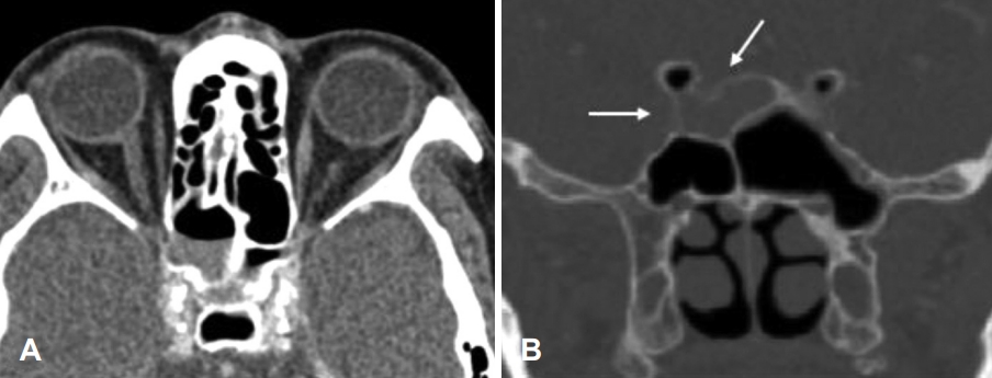
On the third day of hospitalization, the radiology department requested to collaborate with the otolaryngology department about the lesion on the Onodi cell. No rhinologic symptoms such as rhinorrhea, nasal obstruction, or hyposmia were reported; the endoscopic examination showed slightly swollen mucosa without purulent rhinorrhea. On the fourth day of hospitalization, paranasal sinus CT was taken. Compared with the MRI taken 3 days before, the lesion filled with Onodi cells and ACP was reduced, and there were erosions in the Onodi cell walls but no bony defect (Fig. 3). It was thought that the sinus lesion caused the nerve palsy, so an intravenous (IV) antibiotic injection (levofloxacin 500 mg qd) was started, and the emergent operation was performed on the same day.
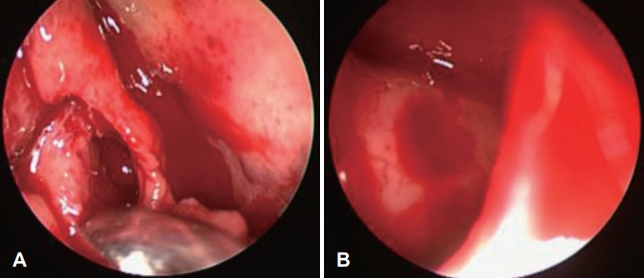
Under general anesthesia, fronto-maxillo-ethmoidectomy was done. Onodi cells were removed, and the natural foramen was widened sufficiently. Severe edema of the mucosa around the opening of the Onodi cell was found (Fig. 4A). Onodi cells were filled with thick mucus secretion and were removed, and there were no defects in the inner optic nerve canal or cranial base (Fig. 4B). Permanent pathology showed chronic inflammation. After surgery, the patientŌĆÖs discomfort of moving the eyeball decreased, but diplopia still remained. After IV antibiotic maintenance for 3 days, oral antibiotics (cefditoren, roxithromycin for 2 weeks), and steroids (prednisolone 10 mg maintained for 1 month) are maintained after surgery. The patient was discharged on the 5th day after surgery. Periodic follow-up was performed simultaneously at the departments of ophthalmology and otolaryngology. On the 20th day after surgery, ptosis was relieved and exotropia was partially resolved. Therefore, the steroid dose was gradually reduced and maintained for 1 month. At the postoperative 2-month follow-up, previously observed ptosis and exotropia were completely resolved (Fig. 5C), and no sinus lesions were observed on CT (Fig. 5A and B).
Discussion
Considering that the lesions of the Onodi cell and ACP are similar to those of the sphenoid sinus, it can be expected that their pathophysiology will be similar. Because the anatomical barrier of the posterior sinus is weak and the bone layer of the sphenoid sinus is thin, it easily spreads when inflamed, showing various symptoms related to surrounding important structures [6]. In particular, when there is a correlation between cranial and ocular complications, it should be actively treated and not underestimated. Onodi cells are located at the back of the ethmoid sinus cells and are located laterally and superiorly to the sphenoid sinus. During normal growth, ethmoid sinus cells expand into the sphenoid sinus, causing failure of the sinus wall or creating an intracranial connection. Clinically, when the optic nerve and the internal carotid artery are located inside the cells, caution is required during surgery. The prevalence varies greatly, and it has been reported to vary from 12.7% to 47.3% in a study involving Koreans [7]. Although the prevalence of Onodi cell sinusitis is not known in detail, in the case of sinusitis confined to the sphenoid sinus in a similar location, it has been reported to be about 1% to 2.7% of all sinusitis [8].
Although there are cases of pus or postnasal drip in the sphenoethmoidal recess or nasopharynx through endoscopic examination, there are many cases with normal findings. If posterior cell sinusitis is suspected, diagnosis through imaging is required, and CT is the most useful. MRI can be used in cases of decreased visual acuity or cranial nerve palsy or to differentiate other pathologies such as mucocele, benign tumors, cerebral aneurysms, and internal carotid aneurysms [9].
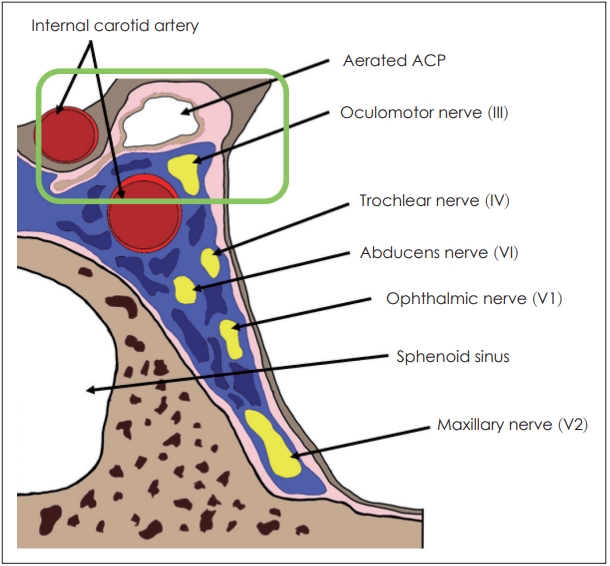
The relationship between sinus disease and cranial nerve palsy is frequently observed in neoplastic and malignant tumors, but it has been reported in 12% of sinus inflammatory diseases [10]. When looking at cases of cranial nerve palsy related to sphenoid sinusitis, the most common was the 6th nerve palsy, followed by the 3rd nerve palsy [11]. The reason is thought to be that the 6th nerve is long and is located closest to the sphenoid sinus on the medial side of the cavernous sinus [2]. However, it is the 3rd nerve, not the 6th nerve, that runs closest to the ACP and the cavernous sinus. Hopf-Jensen, et al. [12] reported a case of 3rd nerve compression by a mucocele of the ACP. They believed that the oculomotor nerve palsy arose because the 3rd nerve was closest to the mucocele. And in our case, the symptoms arose because of the compression of the 3rd nerve due to inflammation of the ACP. In the cavernous sinus, the 3rd nerve passes closest to the inferolateral side of the ACP, so it is thought that it will be invaded more easily than other extraocular cranial nerves when the ACP is inflamed (Fig. 6) [13].
Treatment to prevent complications of sinusitis is broadspectrum antibiotic therapy. Surgery should be considered if there is no improvement or worsening within 24 to 48 hours or if diagnosed as fungal or mucocele. Even when the neurological symptom occurs, immediate surgery is required to restore nerve function [9]. Moreover, active drainage should be considered if a sinus lesion is found in a patient with ocular symptoms. In our patient, the Onodi cells were inflamed in the ACP, and 3rd nerve palsy occurred. MRI confirmed partial compression of the 3rd nerve in the cavernous sinus. As a mechanism of nerve paralysis, it is thought that temporary compression of the 3rd nerve caused the symptoms by direct compression due to sinus lesions or inflammatory edema change to surrounding tissues through bone erosion.
The main causes of isolated 3rd nerve palsy include microvascular ischemia, trauma, compression due to mass and iatrogenic [5]. As there are various causes, treatment must correct each cause, and the overall recovery rate is 48.3%-70.3%. With the emergent surgical drainage, using systemic steroids is important in cranial nerve palsy because it relieves symptoms by regulating the substances related to the immune response and reducing edema near the nerves [14]. It took quite a while to recover completely in this case. Possible causes are as follows: First, early intervention was delayed because the possibility of sinusitis was overlooked. The optimal treatment time for isolated 3rd nerve palsy is not known. However, El Mograbi and Soudry [11] analyzed 17 cases of isolated ocular nerve palsy caused by sphenoid sinusitis, and reported that surgery was performed within an average of 2.5 days after symptom onset, and complete recovery was usually achieved within 1 month [11]. In our patient, surgical treatment was performed 1 week after the onset of symptoms, and the recovery period was 2 months. We believe that the rather late treatment may have affected the recovery period. Second, surrounding cytokine release with edematous mucosa around the nerve can delay relieving cranial nerve symptoms.
In this case, we treated a patient who developed isolated 3rd nerve palsy due to inflammation of the Onodi cell and APC found on MRI. Since cranial nerve palsy is not a common complication of sinusitis, if it is overlooked, the diagnosis may be delayed, leaving fatal complications. Considering various symptoms, recognition through imaging tests for early diagnosis and rapid and active treatment will have an important influence in determining treatment policy.