Introduction
Mandibular reconstruction using fibular free flap is nowadays considered the gold standard method to reconstruct the mandibular defect [1]. Mandibular reconstruction is always essential and difficult in terms of functional and esthetic restoration of mandible. The purpose of reconstruction of oral cavity and mandible especially is not only to restore the anatomy but also to recovery the speech, swallowing and facial contours. Therefore, the success of reconstruction affects the quality of patientвҖҷs life and social activities. The traditional freehand technique of mandible reconstruction requires a precise bone works to restore the U-shaped curvature of mandible. However, it is a very difficult to determine the exact location and angle of the cutting section of the fibula dependent solely on surgeonвҖҷs experience. In the past, mandibular reconstruction has been performed depending only on the surgeonвҖҷs experience without an accurate surgical plan. This has resulted in additional osteotomy and alternation of surgical plan during operation and eventually to prolonged ischemic times and improper mandibular reconstruction. Recently, from the perspective of personalized and functional reconstruction, several software programs that can make accurate preoperative surgical plans in a three dimensional (3D)-virtual system have been developed and used to improve this problem. This software and surgical technique have been called in various terms including digital surgery, computer-aided design, computeraided manufacturing, 3D printing technology and virtual surgical planning (VSP) [2,3]. In recent years, we have used the VSP to assist the mandibular reconstruction and achieved excellent clinical outcomes. The aim of this study is to describe our experience and to share the tips and pitfalls of mandibular reconstruction with VSP.
Subjects and Methods
Patients
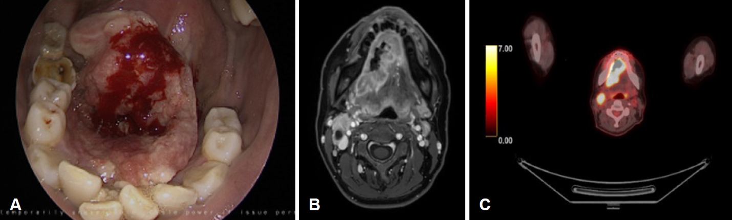
A 45-year-old female with a history of smoking initially presented with painful ulcerative mass at right mouth floor that started 3 months ago (Fig. 1A). This patient was diagnosed with squamous cell carcinoma of mouth floor right (cT4aN2cM0) by punch biopsy and imaging work up. Neck CT and MRI showed a huge irregular mass in the mouth floor and anterior tongue areas. And no distant metastasis was found on PET-CT (Fig. 1B and C). This patient was especially suspected of invading the mandible cortex by MRI and dental scan imaging. We decided to perform a wide excision of mouth floor and anterior tongue with subtotal mandibulectomy, modified radical neck dissection bilateral, tracheostomy and reconstruction with fibular free flap with VSP. Written informed consent was obtained from this patient before surgery. This study was approved by the Institutional Review Board of Yongin Severance Hospital.
VSP
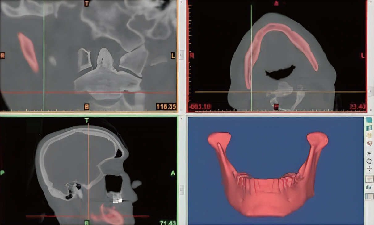
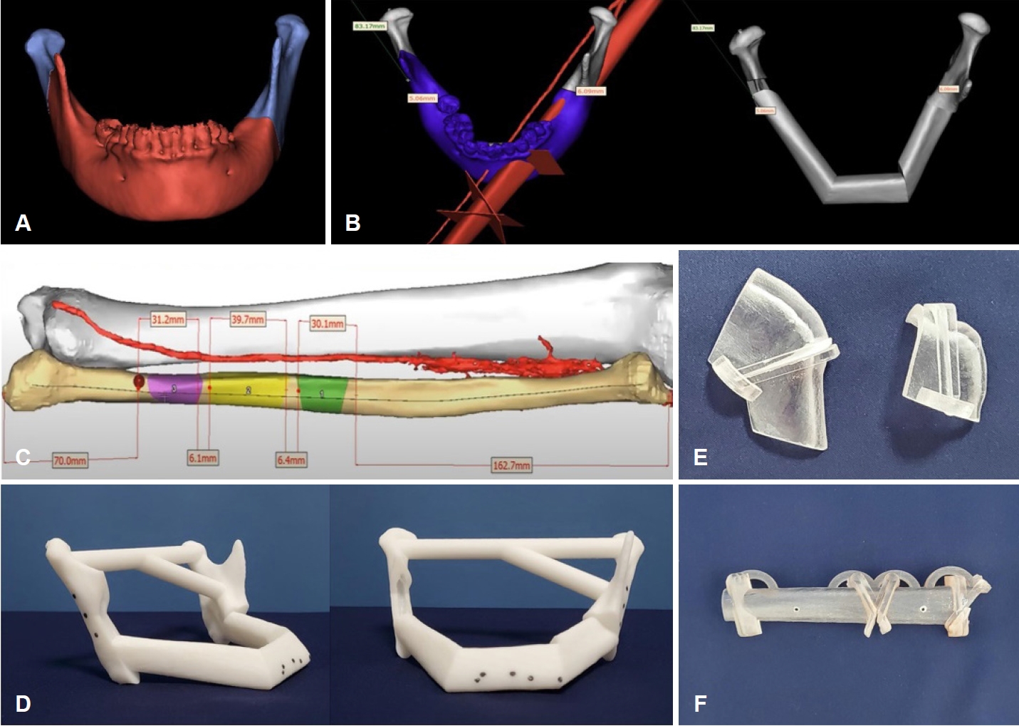
We used the Mimics Innovations Suite software program (Ver.23; Materialise, Leuven, Belgium) for VSP. This technique began with the collecting of high resolution-angiographic CT scan of the patientвҖҷs facial bone and lower extremity. The acquired digital imaging and communications in medicine (DICOM) data format from thin-section (1.5 mm) high resolution CT were sent to the 3D modeling company to create a 3D images (Fig. 2). We were able to determine the oncological safe surgical 2 cm-margin of subtotal segmental mandiblectomy using the created 3D reconstruction mandible images before the surgery (Fig. 3A). The next step is the merging of 3D reconstructed mandible and fibula images to determine the design and molding of the fibular bone flap according to the virtual resection of tumor margin (Fig. 3B). We could simulate the fibular bone flap to restore the pre-mandibulectomy shape of the mandible according to the range and location of the defect. And the length and angle of a three-segment fibular bone graft was virtually designed according to defect size (Fig. 3C). Based on these results, we could create 3D models including neo-mandible, mandiblular and fibular cutting guides through sending the VSP data to the modeling company (Fig. 3D-F). Then, the titanium plate was pre-bent according to the 3D neo-mandible models. The 3D models could finally transform the preoperative virtual design into the actual operation device. This process usually took about 7 days. This imaging was used to explain and communicate the patientвҖҷs current condition, extent of lesions and purpose and method of surgery. The patients with assistance of VSP have an additional surgical costs.
Segmental mandibulectomy
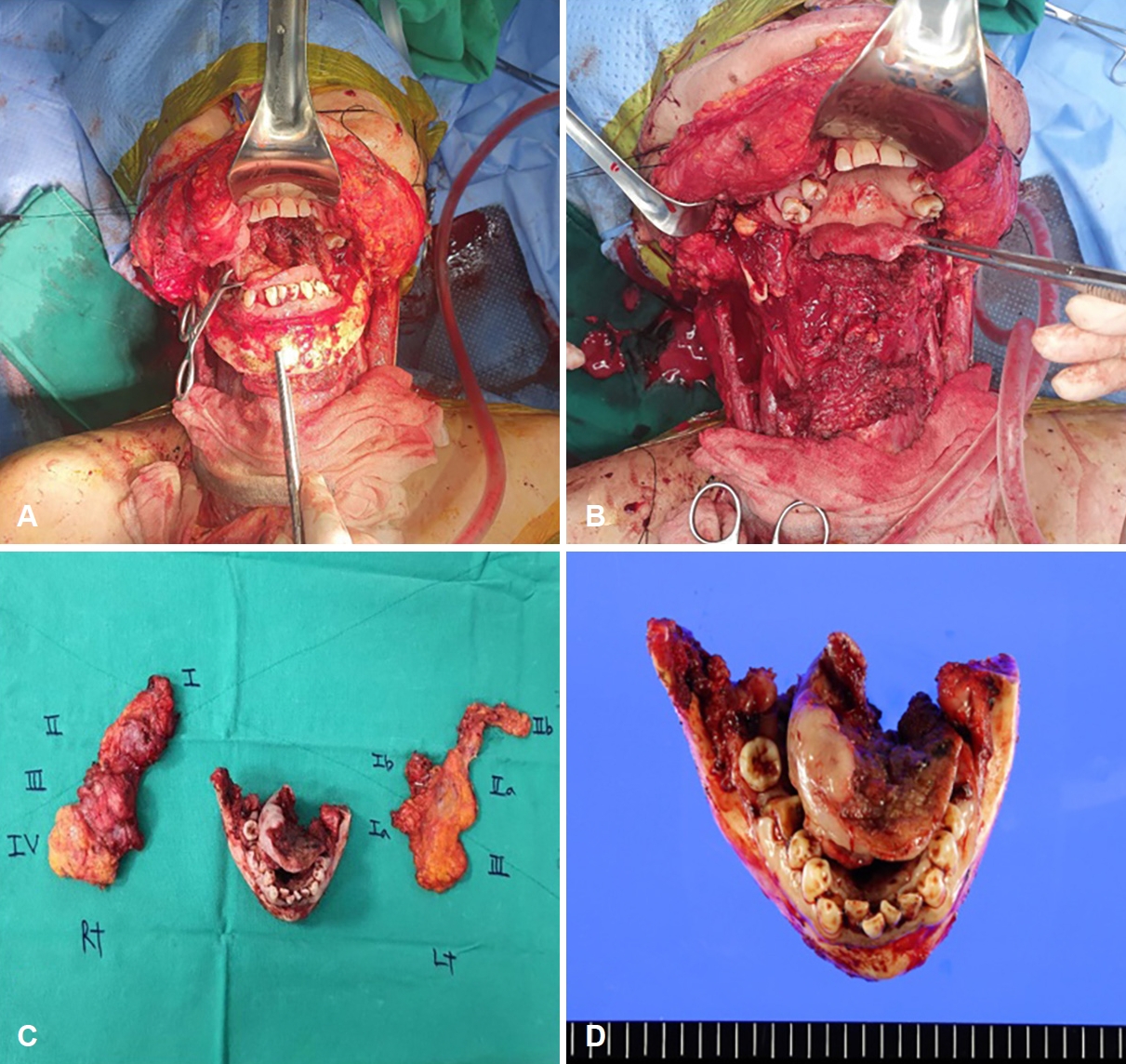
Under general anesthesia, we firstly performed a tracheostomy. And then, we performed a modified radical neck dissection bilateral and segmental mandibulectomy from mandible ramus right to mandible angle left with wide resection of a mouth floor and anterior tongue under the guidance a pre-made mandibular cutting guides by the use of oscillating saw (Fig. 4).
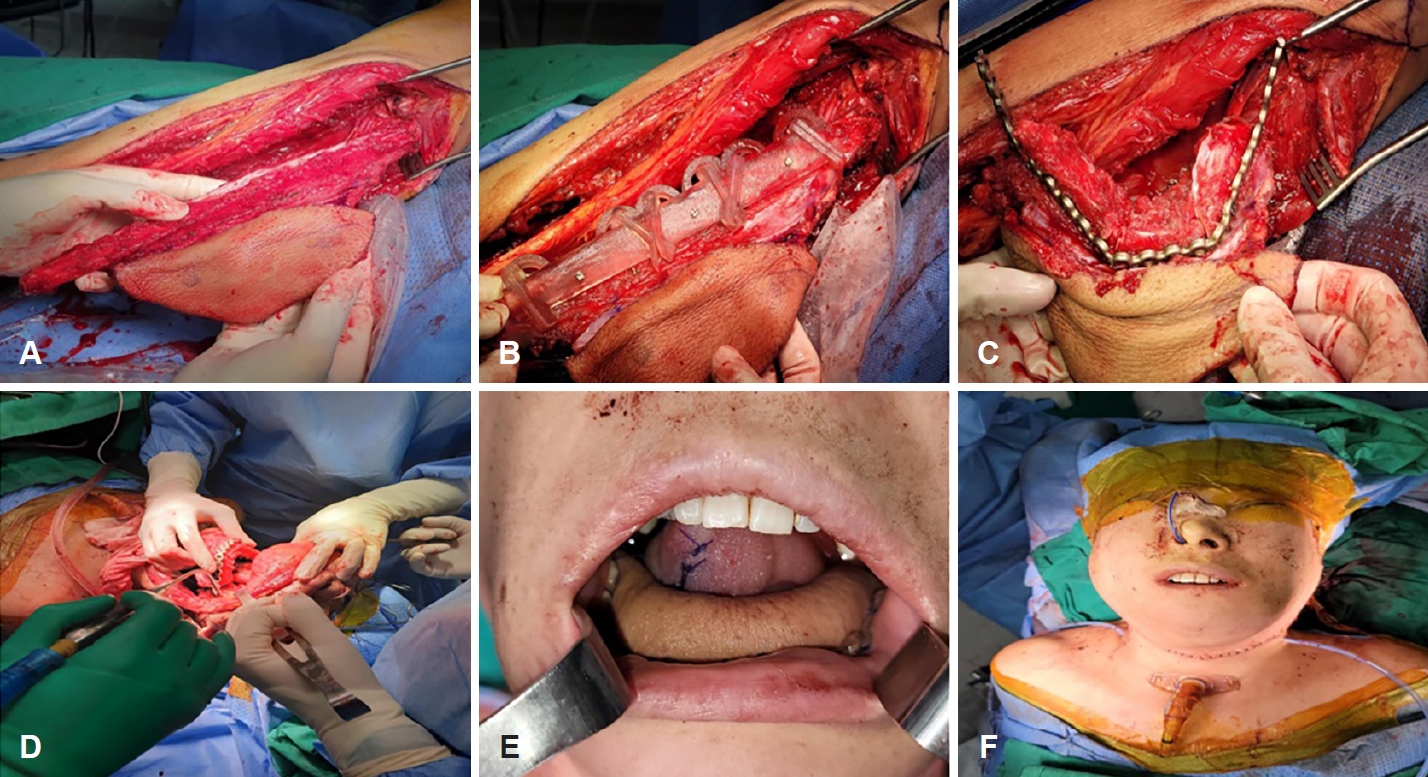
Fibular flap harvest and insetting
Left fibular free flap harvest began in the same technique method as conventional and three-segment fibular osteotomies were performed using pre-made fibular cutting guide. And then, neo-mandible was made with fixation of pre-bent titanium plate before clamping the pedicle. Insetting of neomandible and vascular anastomosis was performed in the same manner as in the conventional methods (Fig. 5).
Postoperative care
A soft fluid diet through the nasogastric tube started after the operation, and the oral diet was completely consumed after 3 weeks. Decannulation was performed 10 days after operation. The patient was discharged from the 32 days after the operation followed with adjuvant chemo-radiotherapy. The dental implant restoration is planned in about a year.
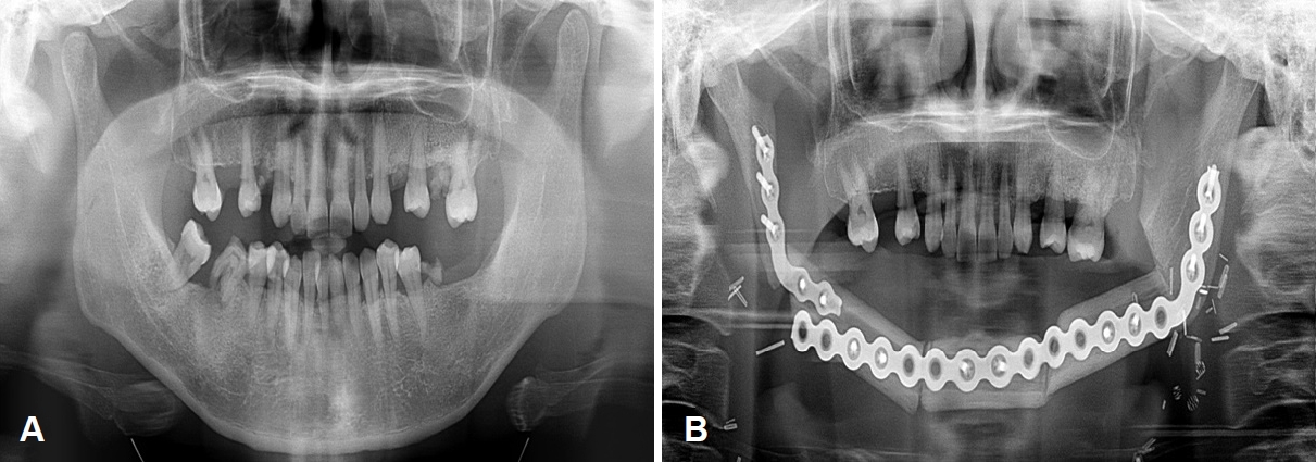
Results
The mean total ischemic (the time from vascular pedicle clamping to the completion of vascular anastomosis) and operation time were 102 and 621 minutes. No postoperative complications including flap congestion or necrosis, infection, orocutaneous fistula, hematoma were observed. During follow-up period (13 months), no recurrence was observed by physical examination and imaging work up and the patient was able to have a proper mouth opening and eat a soft diet (Fig. 6). And the patient was very satisfied with postoperatively facial contour.
Discussion
The mandibular reconstruction is always a highly challenging work because it is very hard to fully and exactly restore the curvature and shape of the mandible. It is also very difficult to improve the accuracy and efficiency of free flap reconstructive surgery that rely solely on the experience of surgeon. As personalized and precision medicine advances, VSP has been developed and adopted in head and neck reconstructive surgery to help surgeons by providing a preoperatively 3D model images and cutting guides. The application of VSP in mandible reconstruction has made it possible to perform an accurate osteotomy through preoperative 3D surgical plan and to diminish the ischemic time obviously. In real operation, we could experience in shorter operation time than a conventional procedure. And flap ischemic time was also significantly reduced compared to traditional technique. Several studies have also reported the benefit of operative time [4,5]. Use of VSP could also offer patients with accurate, effective visual communication and counselling before the surgery. And it could determine the safe and wide range of surgical margin because the surgeon could observe the surrounding structures by 3D reconstruction technology. However, despite its various advantages, it also has some disadvantages. Pre-made cutting guides for mandibulectomy could be sometimes too smaller than initially expected during real surgery. Because, the tumor may have grown in size from when VSP was initially manufactured [6]. As a result, mandibular guides become a good for nothing, it can cause confusion for surgeon during surgery.
Although there are potential disadvantages with VSP, we believe that these is fully modifiable and have significant advantages including savings in operative time and accuracy of flap surgery. Therefore, the using of 3D VSP in mandibular reconstruction is considered to improve the functional and aesthetic outcomes by enabling precise resection and reconstruction.