Introduction
Foreign body (FB) inhalation/ingestion is common among pediatric patients, and early identification followed by prompt management is crucial to minimize morbidity [1]. The introduction of various magnet toys has increased the incidence of pediatric magnet ingestion [2]. Ingestion of multiple magnets is particularly hazardous because magnetic attraction may lead to pressure necrosis in vital organs.
We report a case of secondary tracheoesophageal fistula (TEF) following ingestion of multiple magnets in a 3-year-old girl.
Case
A previously healthy 3-year-old girl was referred to our pediatric emergency room after having ingested multiple magnet beads. The exact time of ingestion was uncertain because the event was not witnessed by her parents, but was presumed to be 8-9 days earlier while playing with her magnet bead toys. The parents initially took the patient to a nearby hospital when she developed a wheeze 2 days before; radiographs showed multiple FBs in the trachea, esophagus, and small bowel.
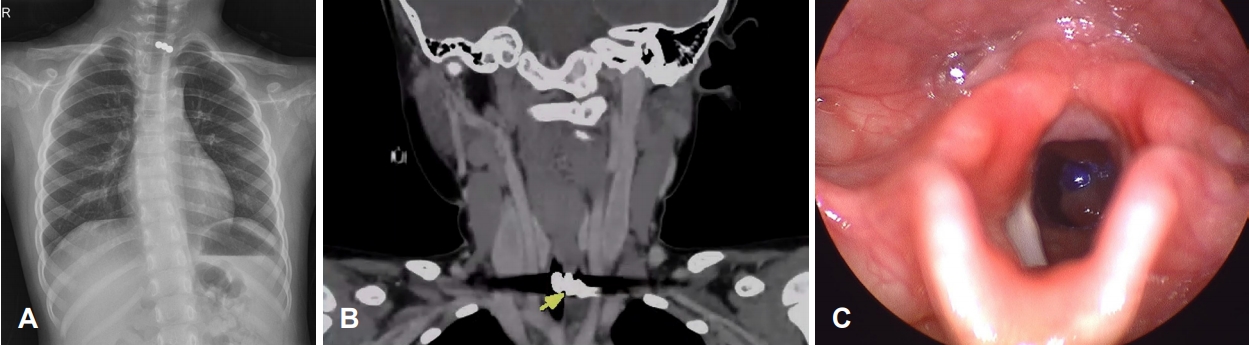
At the time of presentation, the child was healthy without any signs of respiratory distress, dysphagia, or lethargy. The physical examination was unremarkable. The posteroanterior chest radiograph (Fig. 1A) and abdominal radiograph showed three metallic FBs in the lower neck and aggregated metallic FBs in the lower-middle abdomen. Neck computed tomography (CT) (Fig. 1B) scanned for detailed evaluation of the ingested beads, revealed one FB in the trachea and the other two in the esophagus. Upon laryngoscopy (Fig. 1C), the tracheal FB was visible in the proximal trachea, consistent with the CT findings.
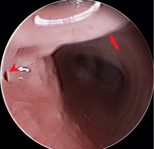
Emergency suspension laryngoscopy was performed under intravenous anesthesia with spontaneous respiration and high flow nasal oxygen (STRIVE Hi) [3,4] at 40 L/min and FiO2 of 1.0. The tracheal FB was located just below the cricoid cartilage and was surrounded by granulation tissue (Fig. 2A). The tracheal FB was successfully removed using magnetic attraction toward a pair of metallic upward forceps (Fig. 2B). An esophagoscope was introduced and the remaining two magnetic beads were removed in the same manner using optical forceps with KILLIAN bean jaws (KARL STORZ, Tuttlingen, Germany) (Fig. 2C). A tiny TEF was visible beneath the tracheal granulation tissue (Fig. 3). The granulation was left intentionally untouched to promote spontaneous healing of the fistula. Since the fistula was located at the optimal position of an endotracheal tube tip, the laryngeal mask airway (LMA) was intubated before proceeding to exploratory laparotomy. A total of 14 magnet beads were extracted from the small bowel; four and ten beads were extracted from the distal and proximal loops, respectively. Two enteroenteric fistulae were identified; one was primarily repaired, whereas segmental resection with enteroenteric anastomosis was performed for the other.
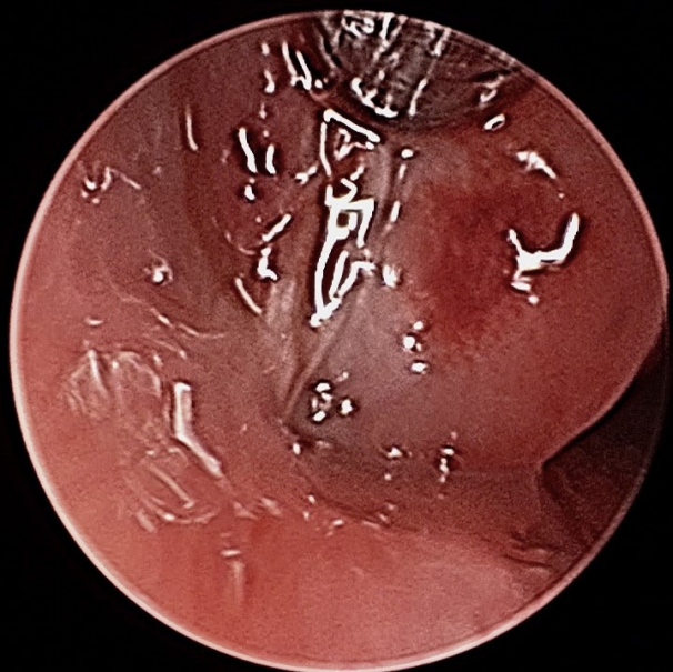
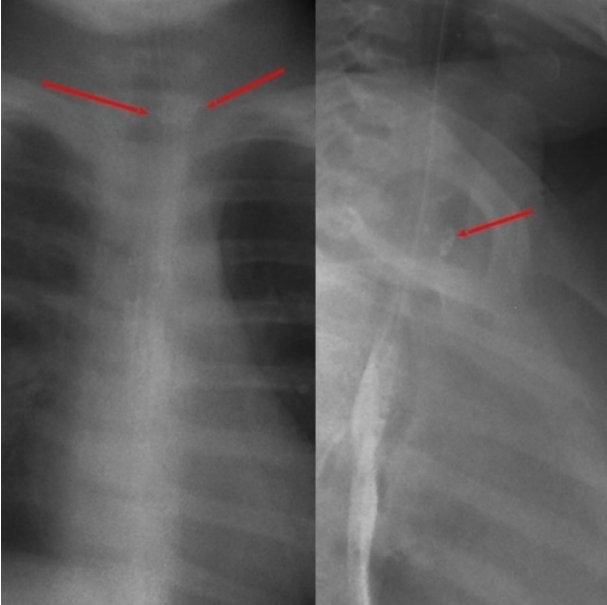
The patient was kept nil per os (NPO) for a week. A follow-up suspension examination on postoperative day 7 showed a well-healed TEF and decreased granulation tissue (Fig. 4). Esophagography performed on postoperative day 10 showed a small outpouching lesion in the anterior aspect of the upper esophagus, but no definite leakage (Fig. 5). After 2 months of Levin tube feeding, the patient was successfully converted to oral feeding without any complications.
Discussion
A vast majority of FB aspiration cases occur unwitnessed and remain asymptomatic, leading to delayed diagnosis and sometimes fatal consequences [5]. Treatment plans may differ depending on the FB characteristics, time of ingestion, severity of symptoms, and radiographic findings [2]. However, in cases involving multiple magnets, there is a high risk of interaction between the magnets and complications such as enteroenteric fistulae, bowel perforation, and intestinal necrosis have been reported [6,7].
The present case was unique because of the simultaneous ingestion and aspiration of magnetic beads. In this complex situation, several factors were taken into consideration during treatment planning.
From the anesthetic perspective, intubation was not a good option because the FB was visible just below the vocal cords. The STRIVE Hi technique [3] was used instead, and provided uninterrupted access to the airway while facilitating oxygenation. Furthermore, the LMA was intubated before proceeding to abdominal surgery to prevent exacerbation of the TEF.
Another factor was the shape and location of the FBs. The metallic bead in the proximal trachea made it difficult to use a rigid bronchoscope. Instead, a suspension laryngoscope was used to identify and remove the tracheal FB. Moreover, spherical FBs like magnet beads are challenging to extract. Attempts to grasp the FB may instead push it distally. Dexterous handling of an appropriate instrument is the key to successful retrieval. Based on the unique properties of magnets, metallic instruments were selected; upward forceps were used to swipe the metallic bead distal to the proximal trachea and prevent it from falling backwards during detachment.
During retrieval, the magnetic interaction between the FBs in the esophagus and trachea is lost, which may cause the remaining FB to migrate distally. Therefore, the tracheal FB was retrieved first to prevent airway obstruction and acute desaturation. Conversely, a FB remaining in the esophagus is much less of a threat and may be accessed through rigid esophagoscopy or esophagogastroduodenoscopy.
Based on the time of ingestion and preoperative radiographic findings, a secondary TEF was suspected, and tracheal resection with end-to-end anastomosis was planned for the worst-case scenario. Fortunately, the TEF was small and completely covered by tracheal granulation tissue; it was thus allowed to heal secondarily. After a week of NPO with Levin tube feeding and intravenous administration of proton pump inhibitors, the fistula resolved spontaneously.
Although the present case involved successful removal of multiple magnets, such cases often have poor outcomes. Despite warning labels on magnetic toys, the risk of their misuse by children is often understated. The potential risks associated with metallic toys need nationwide recognition to minimize accidents.
In conclusion, we report a unique case of multiple magnets in the aerodigestive tract. Thorough understanding of the features of magnetic FBs is the key to successful management. With the introduction of mini-magnet home supplies and toys, magnet-related emergencies can no longer be neglected. Parents should be aware of the potential risk, and health care providers should give due importance to the proper management.