양성종양으로 오인된 혀기저부의 유리화 투명세포암종: 증례 보고
Hyalinizing Clear Cell Carcinoma of the Base of Tongue Mistaken for Benign Lesion: A Case Report
Article information

Trans Abstract
Hyalinizing clear cell carcinoma (HCCC) is a rare, low-grade malignant tumor of the salivary gland. It usually originates from the minor salivary gland, with the most common site being the palate, followed by the lips and the buccal mucosa. The occurrence of HCCC at the base of the tongue (BOT) is extremely uncommon, thus it must be differentiated from other malignant clear cell tumors. Immunohistochemistry is a useful tool to make an appropriate diagnosis. To obtain the best prognosis for HCCC, complete surgical resection is necessary. Here we report a case of a 47-year-old male with a benign-looking neoplasm in the right BOT, presenting with throat discomfort. A simple excisional biopsy revealed proliferative nests of clear cells within a hyalinized fibrous connective tissue. The final diagnosis by immunohistochemistry was HCCC with a positive resection margin. Re-operation secured a safety margin, and the lesion was completely resected.
서 론
유리질 투명세포암종(hyalinizing clear cell carcinoma, HCCC)은 매우 드문 부침샘 악성종양으로, 전체 침샘 종양의 1% 미만으로 발생한다고 보고되고 있다[1]. 암종의 호발 부위는 경구개이고 다음으로 입술과 구강 점막 순으로 발생하며, 구인두에서의 발생은 매우 드문 것으로 알려져 있다[1]. 암종의 기질이 광학현미경상에서 투명하게 보이는 것이 특징이며, Milchgrub 등[2]에 의해 1994년 처음으로 명명되었다. 대부분 저악성도 암종으로 점막하층에서 잘 발생하며, 크기가 커지기 전에 환자들이 증상을 느끼는 경우는 드물지만, 전이를 한 증례도 보고된 바 있다[3]. 저자들은 구인두 불편감을 주소로 내원한 47세 남자 환자에서 설 기저부의 양성종양으로 오인된 유리질 투명세포암종을 경험하였기에, 문헌 고찰과 함께 증례 보고를 하고자 한다.
증 례
47세 남자 환자가 1년 전부터 시작된 구인두 불편감을 주소로 내원하였다. 목소리 변화나 호흡곤란 등의 다른 증상은 없었으며 타 동반 질환도 없었다. 신체 진찰에서 우측 설 기저부에 주변과 경계가 비교적 명확한 둥근 모양의 양성종양으로 보이는 종괴가 관찰되었고(Fig. 1), 목에서 만져지는 결절은 없었다. 촬영한 CT에서 우측 설 기저부에 반응성 림프구 증식을 시사하는 2×2 cm 크기의 종괴가 관찰되었고(Fig. 2), 다른 특이소견은 없었다. 양성종양이라 판단하여 진단을 위한 예비 생검 없이 단순 절제를 계획하였다. 전신마취하 설기저부의 종괴를 전 절제하였고, 조직 병리 결과 저악성도 암종 가능성이 높다고 진단되어 면역조직화학 검사를 진행하였다. 결과는 cytokeratin, p63 양성, carcinoemyronic antigen, S-100 음성으로 나타났다(Fig. 3). 따라서 다른 부침샘 종양들과 구별해야 하는 전이성 투명세포암종은 배제되어 유리질 투명세포암종으로 최종 진단되었다. 절제면이 양성이었기 때문에 이후 환자는 전이를 배제하기 위해 PET-CT를 시행하고, 수술 부위 이외에 특이소견이 없어 음성 절제면을 얻기 위해 재수술을 시행하였고 병변은 완전히 절제되었다. 재시행한 조직학적 검사에서는 0.5 cm의 안전 절제연을 확보하여서 추가 치료는 시행하지 않았고, 환자는 현재 수술 후 5개월 국소재발 없이 외래에서 추적 관찰 중인 상태이다(Fig. 4).
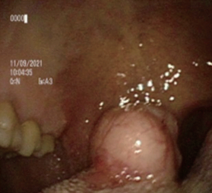
Right tongue base showing benign-looking 2×2 cm sized mass. There was no surface ulcer, nor inflammation.

Neck CT, 2×2 cm sized mass-like lesion in tongue base, suggesting reactive lymph node. There was no other abnormality in CT. A: Axial view. B: Coronal view.
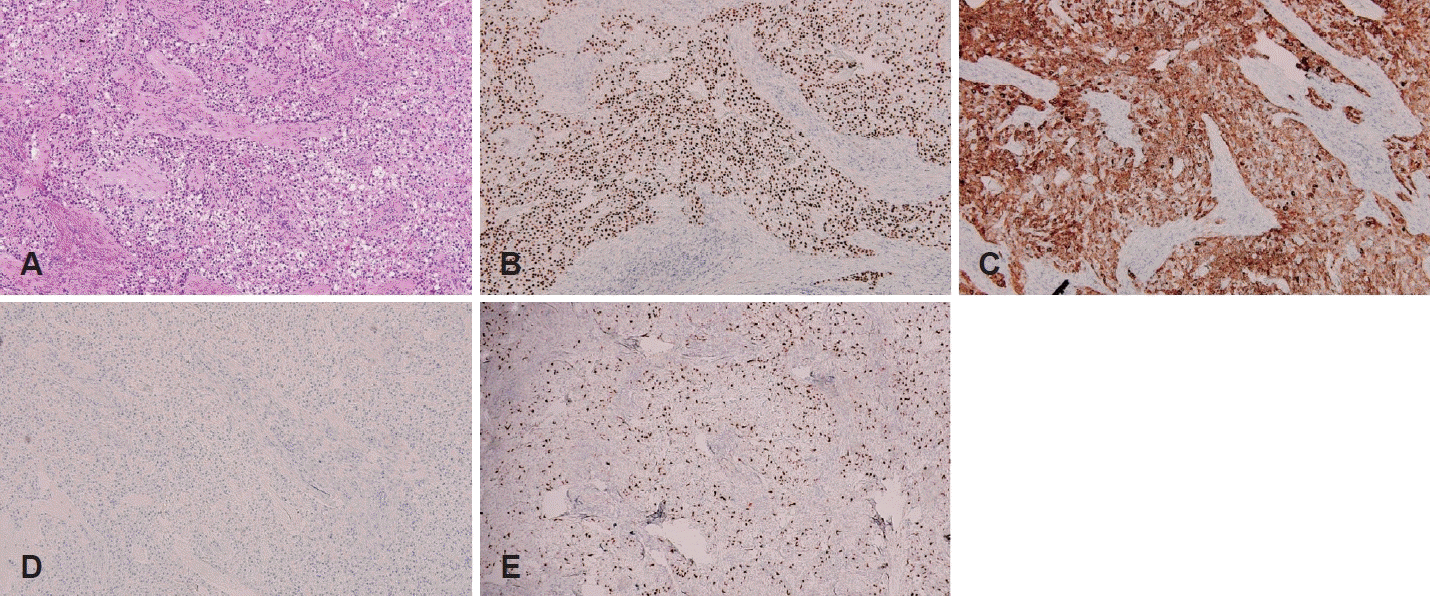
Histopathology of the main mass. A: H&E stain (×20), the tumor consists of islands, cords, trabeculae, and a sheet of neoplastic epithelial cells. B: Immunohistochemistry, positive for p63 (×20). C: Positive for cytokeratin (×20). D: Negative for carcinoembryonic antigen (×20). E: negative for S-100 (×20).

Right tongue base, 5 months after operation. There was no sign of recurrence.
고 찰
유리질 투명세포암종(HCCC)은 드문 타액선 종양으로 전체 타액선 종양의 1% 미만을 차지한다[4]. 유리질 투명세포암종은 편평 분화가 있는 모든 침샘 부위에 나타날 수 있지만 대부분 경구개, 입술 등에 발생하며, 설기저부에는 매우 드물게 발생하는 것으로 알려져 있다. 일반적으로 유리질 투명세포암종은 표면에 궤양이 생기지 않으며, 천천히 성장하고, 통증이 없는 점막하 종괴로 나타난다[3]. 따라서 환자는 발병 후 몇 개월이 지난 후 불편감 등의 증상을 나타내며 악성으로 의심되는 임상 특징을 보이지 않아 양성 질환으로 오인할 수 있다. 유리질 투명세포암종은 적절한 치료 및 관리를 위해 부침샘에서 발생할 수 있는 다른 공격적인 종양과 구별되어야 하며, 조직학적 검사, 특히 면역염색으로 구별이 가능하다[2,5,6].
구별해야 할 질환으로는 투명세포 신암종(renal cell carcinoma), 상피-근상피 암종(epithelial-myoepithelial carcinoma), 점막표피양 암종(mucoepidermoid carcinoma), 포상세포암종(acinar cell carcinoma) 및 투명세포 치성 암종(clear cell odontogenic carcinoma) 등이 있다[1]. 유리질 투명 세포암종은 p63 및 cytokeratin (CK5, CK7, CK8, CK14 및 CK19)에 대한 면역 조직 염색에 양성이고 calponin, S-100, smooth muscle actin (SMA) 및 glial fibrillary acidic protein에 대해 음성이다. 또한 Periodic acidic Schiff에 대해 양성이고 mucicarmine에 대해 음성으로 염색된다[3]. 가장 흔한 악성 침생종인 상피-근상피 암종은 유일하게 근상피 표지자(SMA, S-100)에 대해 양성으로 구별된다[3]. 점막표피양 암종은 mucin에서 양성을 나타내어 구별된다. 포상세포암종은 SMA에서 양성을 나타내어 유리질 투명세포암종과 구별되며, 투명세포 치성 암종은 CK에서 염색되지만, S-100이 양성으로 염색되어 감별이 가능하다. CD10은 유일하게 투명세포 신암종에서만 염색되어 유리질 투명세포암종과 감별진단이 가능하다[6]. 그리고 현재 연구에 따르면 면역 조직 염색으로도 감별진단이 어려울 경우, 형광동소분합법(fluorescence in situ hybridization)을 통한 EWSR1-ATF1 유전자의 융합 검출이 유리질 투명세포암종의 확인에 도움이 될 수 있다고 알려져 있다[5,7].
유리질 투명세포암종은 일반적으로 악성도가 낮으며 심각한 질병 관련 사망률과 관련이 없다고 알려져 있다[3,7]. 그러나 어떤 사례에서는 종괴가 후두 입구를 가리며 기도가 좁아져 기관절개술이 필요한 경우도 있었고, 전이성 국소 림프절이 있어 경부림프절 절제술이 필요한 경우와 완화 항암치료가 필요한 일부 드문 증례가 보고되었다[1,3,6,8-11]. 아직 암 병기를 기반으로 한 치료 요법은 정립되지 않았다[2]. 따라서 많은 사례에서 암종의 임상적 특성에 따라 치료 요법이 결정되었다. Oliver 등은 50% 이상의 경우, 치료로 광범위한 국소절제만을 시행하였고, 25.7%에서 고악성도를 보이거나 림프절 전이가 있는 경우 술후 방사선 치료를 하였다고 보고하였다[6,9].
일반적으로 유리질 투명세포암종은 관찰되어도 환자의 불편감이 없을 수 있으므로 양성으로 보이는 종양이 구강 내에서 처음 발견되었을 때 반드시 악성에 대한 가능성을 생각해야 한다. 국내에서는 위 환자의 증례가 혀의 기저부에 나타난 유리질 투명세포암종의 첫 보고이다. 저자들은 양성종양으로 오인할 수 있는 매우 드문 유리질 투명세포암종의 진단과 치료를 경험하였기에 문헌 고찰과 함께 보고하는 바이다.
Acknowledgements
This work was supported by research fund of Chungnam National University.
Notes
Author Contribution
Conceptualization: Min Gyu Kim, Seong Jun Moon, Bon Seok Koo. Data curation: Min Gyu Kim, Da Beom Heo, Seong Jun Moon. Project administration: Bon Seok Koo. Supervision: Bon Seok Koo. Writing—Original draft: Min Gyu Kim. Writing—review & editing: Bon Seok Koo.