이관 풍선 확장술 중 발생한 부정맥 2예: 기전과 치료
Two Cases of Intra-Operative Cardiac Arrhythmia During Balloon Eustachian Tuboplasty: The Mechanism and Treatment
Article information
Trans Abstract
Balloon eustachian tuboplasty (BET), a surgical technique to expand the cartilaginous portion of the eustachian tube by ballooning via opening at the nasopharynx, has been introduced as a useful surgical modality for eustachian tube dysfunction patients. Although BET is known as a relatively safe procedure, we recently have experienced two cases of cardiac complications during balloon inflation. In one case, an asystole occurred for 13 seconds during this procedure; the heart rate was recovered after balloon deflation with an intravenous injection of glycopyrrolate and atropine. In the other case, bradycardia occurred and continued during BET. Heart rate was recovered immediately after deflation of balloon without drug injection. As far as we know, this is the first report of cardiac complications during BET, probably related with trigemino-cardiac reflex. In both cases, no other sequelae remained after the surgery. We report these two cases of cardiac complications that occurred during BET along with a review of literature.
서 론
이관이 폐쇄된 상태로 열리지 않는 폐쇄성 이관 장애(dilatory eustachian tube dysfunction)의 경우 지속적인 이관의 음압으로 인해 삼출성 중이염, 유착성 중이염, 진주종 등의 원인이 될 수 있다[1]. 폐쇄성 이관 장애를 해결하기 위해 Schröder 등[2]은 비인두의 이관 입구로 접근하여 풍선 카테터를 거치시키고 압력을 주입하여 이관의 연골부를 확장하는 이관 풍선 확장술(ballon eustachian tuboplasty)을 소개하였다[3]. 이는 최소 침습으로 비교적 간단하게 이관기능장애를 해결할 수 있으며 만성 이관기능장애 환자들에게 효과가 인정되어 최근 널리 사용되고 있다[2].
이관 풍선 확장술은 비교적 안전한 시술로 알려져 있는데, Schröder 등[2]은 1076개 귀에 이관 풍선 확장술을 시행하였을 때 이하선 부위 기종이 발생한 3예와 경미한 출혈 외에는 심각한 합병증이 없었다고 하였다. 이관 풍선 확장술에 대한 메타분석에서도 신경통과 출혈 외에는 심각한 합병증은 보고되지 않았다[4]. 그러나 저자들은 최근 이관 풍선 확장술을 시행하던 중 이관 내 풍선 카테터를 거치한 뒤 풍선을 부풀리는 순간 발생한 1예의 심장 무수축, 1예의 서맥을 경험하였다. 무수축과 서맥은 현재까지 이관 풍선 확장술의 합병증으로 알려진 바 없기에 증례 보고와 함께 고찰하고자 한다.
증 례
증례 1
양쪽 고막 환기관 삽입술 과거력이 있는 33세 여자 환자가 1주 전부터 시작된 좌측 이통을 주소로 내원하였다. 수술 전 시행한 검사상 좌측 고막의 천공, 좌측귀의 경도의 전음성 난청이 확인되었다. 측두골 CT에서 중이 및 유양돌기 내 특이소견이 확인되지 않아 고실성형술을 계획하였다. 양측 고막 환기관 삽입술 기왕력 및 수술 전 발살바법(Valsalva maneuver)에서 양측 모두 이관 개방이 확인되지 않아 이관기능 장애 의심하에 고실성형술과 더불어 양측 이관 풍선 확장술도 함께 계획하였다. 환자의 측두골 CT상 이관과 경동맥 사이 열개 소견은 관찰되지 않았고 과거력상 심혈관계 질환은 없었다.
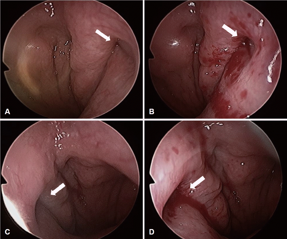
전신마취를 위해 산소포화도, 심전도, 혈압 모니터링하에 프로포폴(propofol) 120 mg을 이용하여 마취를 시행하였으며, 레미펜타닐(remifentanil) 74 mg을 이용하여 마취상태를 유지하였다. 또한 로큐로니움(rocuronium) 50 mg을 근이완제로 사용하였다. 수술은 비강접근법을 통한 좌측 이관 풍선 확장술부터 시작하였다. 비강에 보스민(ephinephrine 1 mg/1 mL)을 적신 거즈를 넣고 3분 후 제거하여 점막을 수축시킨 다음 내시경을 삽입하여 비인두의 이관 개구부를 확인한 뒤 풍선 카테터(Navilloon-e; Mega Medical, Seoul, Korea)를 이관에 삽입하고 12기압까지 풍선 카테터를 팽창시켰다(Fig. 1). 카테터 팽창 전 환자는 정상 심전도를 보였고 맥박수는 분당 80-90회를 유지하고 있었으나 카테터 팽창 직후 환자는 심장 무수축 상태가 되었다. 무수축이 확인된 순간 즉시 카테터의 압력을 빼고 제거하였으며, 글리코피룰레이트(glycopyrrolate) 0.2 mg, 아트로핀(atropine) 0.25 mg을 주입한 뒤 환자의 심장 수축은 정상 맥박수로 돌아왔다. 무수축은 13초 동안 지속되었음을 확인하였고, 이후 정상 심전도로 유지되었다(Fig. 2). 마취과와 상의 후 심전도 모니터링하에 조심스럽게 다시 한번 풍선 카테터를 삽입 후 2분간 풍선 확장술을 시행하였고, 이때는 심박동의 이상 소견은 보이지 않아 확장술 이후 고실성형술까지 진행하였고 수술이 모두 끝날 때까지 환자의 심전도는 더 이상 특이소견을 보이지 않았으며, 수술 후 저산소성 뇌손상 등은 확인되지 않았다.

Pre- and post- operative findings of eustachian tube cartilaginous orifice in case 1. Preoperative left eustachian tube opening at nasopharynx (A, arrow) and postoperative left eustachian tube opening at nasopharynx (B, arrow). Preoperative right eustachian tube opening at nasopharynx (C, arrow) and postoperative right eustachian tube opening at nasopharynx (D, arrow).

Intraoperative electrocardiogram during right eustachian balloon tuboplasty in case 1. Electrocardiogram showed 13 seconds of asystole immediately after balloon inflation at left eustachian tube during balloon eustachian tuboplasty (A). Electrocardiogram showed the recovery of normal rhythm after deflation and injection of atropine and glycopyrrolate (B).
증례 2
반복되는 좌측 중이 진주종으로 11년 전 고실성형술 및 유양돌기 삭개술, 이소골 성형술을 시행했던 과거력이 있는 48세 여자 환자가 진행되는 좌측 난청소견으로 내원하였다. 수술 전 시행한 검사상 좌측 중이 내 재발성 진주종이 의심되는 소견, 좌측 귀의 경도의 전음성 난청 소견이 관찰되었다. 측두골 CT에서도 재발성 진주종 소견이 의심되어 좌측 개방공동 유양돌기 절제술 및 고실성형술을 계획하였다. 또한 수술 전 발살바법 시행 시 양측 고막 모두 반응이 없어 양측 이관 확장술도 함께 계획하였다. 환자의 측두골 CT상 이관과 경동맥 사이 열개 소견은 관찰되지 않았고 과거력상 심혈관계 질환은 없었다.
증례 1과 같은 방법으로 전신마취 후 우측 이관 확장술부터 시행하였고 카테터에 12기압의 압력을 넣자 맥박수가 분당 80-90회에서 35-40회로 감소하였다(Fig. 3A). 마취의와 상의 후 맥박수가 더 이상 저하되지 않아 시술을 지속하기로 하였으며, 팽창을 유지하는 동안 맥박수는 40-50회 정도로 유지되다가 카테터 제거 후 정상으로 회복되었다(Fig. 3B). 좌측에서는 카테터를 평소보다 더 천천히 팽창시켰고, 맥박수 70-80회로 우측 수술 시보다 감소의 정도가 적었고, 시술 중 정상 맥박수로 회복되었다. 이후 고실성형술 및 유양돌기 절제술을 계획대로 시행하였으며 수술 중 및 후에 특이 합병증은 관찰되지 않았다.
고 찰
저자들은 풍선 이관 확장술 중 기존에 보고된 바 없는 2예의 심박동 이상 반응을 경험하여 그 원인에 대해 문헌 고찰을 하였으며, 그 결과 자극 인자인 풍선 확장술의 중단 및 항콜린제와 부교감신경차단제의 사용으로 심박동이 호전된 것에 착안하여 본 현상을 삼차신경심장반사(trigemino-cardiac reflex, TCR)에 의한 현상으로 추측하였다. TCR은 삼차신경의 감각신경분지 자극 시 발생되는 분당 맥박수가 60 이하로 감소, 기저 상태보다 평균동맥압 20% 감소로 정의되는 뇌간 반사로, 삼차신경 감각신경분지 가지들이 기계적인 당겨짐이나 압력등으로 인해 자극되었을 때 심장의 무수축이나, 서맥 등의 부정맥, 저혈압, 무호흡 등의 반응을 유발한다고 알려져 있다[5]. 안과에서는 사시 수술 시 외안근 당김이나 안구 압박에 의해 발생하여 비교적 잘 알려져 있으나[6], 이비인후과적 영역에서는 두개저 수술 중 삼차 신경에 자극에 의해 서맥이 발생한 증례 외에는 잘 알려져 있지 않다[7]. 이관의 감각신경은 삼차신경의 상악분지와 하악분지, 그리고 설인신경에서 기인한 고실신경얼기(tympanic plexus)에 의해 지배되는 것으로 알려져 있는데, 저자들은 이관의 감각을 담당하는 신경들 중 이관의 연골부 감각이 삼차신경의 하악분지에 의해 지배되므로[8,9] 본 증례 환자들에게서 풍선 확장술 중 이관 연골부에 대한 풍선의 압박이 삼차 신경을 자극하여 TCR이 발생했을 것으로 추정하게 되었고, 이는 TCR의 가장 강력한 유발 요인이 기계적인 당겨짐(mechanical stretch)이라고 알려져 있기 때문이다[10,11]. 이 외에도 TCR 발생을 높이는 약물이 소개된 바 있는데, 알려진 약물은 마약성 약제(narcotic agent), 베타차단제(beta blocker), 칼슘채널차단제(calcium channel blocker) 등이다[5,10]. 본 증례 환자들에게도 레미펜타닐(remifentanil) 등의 마약성 약제(narcotic agent)가 흡입 마취 전 사용되었는데, 약제 투여에 의한 교감신경 억제가 vagal tone을 증가시켜 풍선 확장술로 인한 이관 주변 삼차 신경 자극에 의한 TCR 발생에 추가적으로 기여했을 가능성도 있으리라 추측된다.
TCR에 의한 수술 중 합병증을 예방하기 위해서는 삼차 신경의 조작 가능성이 있는 수술 시 앞서 언급한 위험 요인들을 미리 고려해야 할 필요성이 있을 것이다. 수술이 진행되는 동안에도 심전도와 활력 징후를 철저히 감시해야 하며 TCR이 발생하면 즉시 시행 중이던 조작을 중지하여야 한다. 서맥, 저혈압 등이 지속될 경우는 아트로핀(atropine)을 정주하고, 이에도 반응이 없을 경우 에피네프린(ephinephrine)을 투여해야 한다[6]. TCR 발생 예방을 위해 예방적 항콜린약물(anti-cholinergic drug) 사용 또한 고려될 수 있다고 알려져 있다[10,12].
저자들은 최근 이관 풍선 확장술 중 발생한 심장 무수축 및 서맥 합병증 발생 증례를 경험하였기에 원인으로 생각되는 TCR과 대처 방법에 대한 고찰과 함께 보고하는 바이다. 본 증례에서는 전신마취로 수술이 진행되었기에 마취의의 감시하에 심장 합병증이 발생된 즉시 발견하였고 적절한 처치를 시행하여 심장 박동을 정상화 시켰기에 심각한 합병증은 발생하지 않았다. 하지만 본 수술이 국소마취로 외래에서 시행되었을 경우 적절한 대처가 어려웠을 수도 있으리라 생각되었다. 비록 술기 면에서 간단한 이관 풍선 확장술이라 할지라도 가능한 마취과 전문의가 상주하는 수술실에서 심전도와 활력 징후를 잘 모니터링하면서 시행하는 것이 안전할 것으로 생각되며, TCR 발생 가능성을 염두에 두고 가급적 유발 약제 사용을 줄이도록 하고, 확장술 중 심장 서맥, 저혈압 등이 발생했을 경우에는 빠르고 적절한 처치를 통해 심각한 합병증으로 이행되지 않도록 하는 것이 필요함을 제안하고자 한다. 본 증례 보고는 이관 풍선 확장술 중 발생한 TCR에 의한 심장 무수축 및 서맥 등의 심장 관련 합병증으로 기존에 보고된 예가 없었기에 학계 최초로 보고하고자 하며, 본 증례를 통한 저자들의 경험이 향후 이관 풍선 확장술 중 발생할 수 있는 합병증 발생의 예방과 치료에 도움 되기를 바라며, 문헌 고찰과 함께 보고하는 바이다.
Acknowledgements
This study was supported by the Basic Science Research Program through the National Research Foundation of Korea (NRF) funded by the Ministry of Education (2021R1F1A1049965).
Notes
Author Contribution
Conceptualization: Shi Nae Park. Formal analysis: all authors. Methodology: Shi Nae Park. Supervision: Shi Nae Park, Jae Sang Han, Ji Hyung Lim. Validation: Shi Nae Park, Jae Sang Han, Ji Hyung Lim. Visualization: Ji Hyung Lim. Writing—original draft: Junuk Lee. Writing—review & editing: all authors.

