제1형 당뇨병, 소뇌 실조, 진행성 난청을 수반하는 MELAS 증후군 환자 1예
A Case of MELAS Syndrome Involving Diabetes Mellitus, Cerebellar Ataxia and Progressive Sensorineural Hearing Loss
Article information
Trans Abstract
Mitochondrial encephalopathy, lactic acidosis, and stroke-like episodes (MELAS) syndrome is a maternally inherited mitochondrial disorder. MELAS syndrome manifests different clinical symptoms due to mitochondrial heteroplasmy and individual susceptibility and tends to be aggravated by metabolic events. It presents various clinical manifestations including hearing impairment, diabetes mellitus, and stroke like episodes. However, to date, only a few cases of MELAS syndrome accompanied with cerebellar ataxia have been reported in Korea. We hereby present an unusual case of a patient diagnosed with MELAS syndrome involving diabetes mellitus, cerebellar ataxia and sensorineural hearing loss, which were aggravated by metabolic stresses such as pregnancy and hypoglycemic shock.
서 론
Mitochondrial encephalopathy, lactic acidosis, and stroke like episodes (MELAS) 증후군은 가장 흔한 미토콘드리아 질환으로 미토콘드리아 RNA의 3243A→G 변이에 의해 주로 발생한다. 뇌졸중 유사 증상, 기억력저하, 경련, 재발성 두통, 난청, 당뇨병 등이 동반된다[1]. MELAS 환자에서 감각신경성 난청은 75%에서 동반되는 것으로 알려져 있다[2]. 3243A→G의 미토콘드리아 점 돌연변이는 국내보고에서는 비증후군성 감각신경성 난청이 있는 환자 129명 중 1명(0.78%)의 빈도로 확인되었으며[3], 일본에서는 감각신경성 난청 환자 319명 중 1명(0.314%) 빈도로 확인되었다[4]. 반면 난청 가족력이 있는 경우 31명 중 4명(12.9%) 빈도로 3243A→G 미토콘드리아 점 변이가 확인되었다[5]. 이처럼 난청은 MELAS에서 비교적 흔하게 나타나는 증상이지만 소뇌위축이 동반되는 경우는 매우 드물다. 국내 보고된 MELAS 증후군 증례 중 감각신경성 난청 그리고 소뇌위축과 동반되어 자세불안정을 호소하는 경우는 1예뿐이었다[6].
저자들은 제1형 당뇨병과 진행하는 양측성 감각신경성 난청이 있는 환자에게 두통, 어지럼, 자세불안정이 발생한 것을 계기로 분자유전학적 검사와 영상학적 검사를 통하여 소뇌 위축이 동반된 MELAS 증후군을 진단하였고, 환자의 가족력을 통하여 MELAS 증후군이 유전되는 가계를 발견하여 문헌 고찰과 함께 보고하는 바이다.
증 례
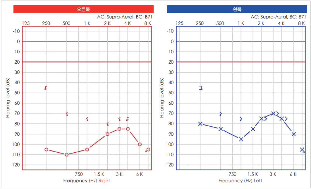
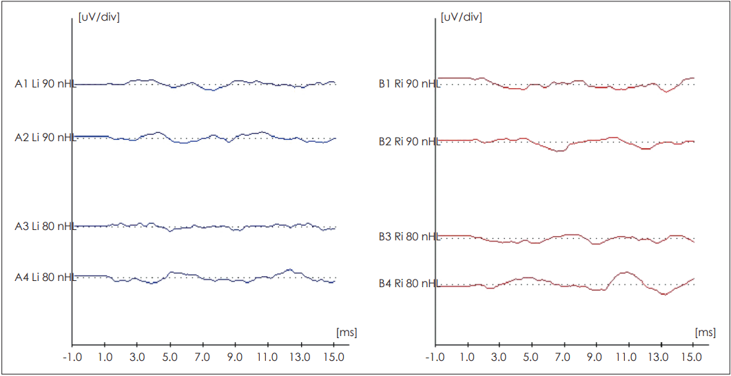
44세 여자가 양측 청력저하와 어지럼으로 내원하였다. 12년 전 두부외상 후 우측 난청이 발생하였고, 8년 전 둘째 아이 임신과 저혈당쇼크 이후 좌측 난청이 발생하였다. 이후 청력은 점점 떨어져 들리지 않았고, 좌측은 보청기를 사용할 수 있는 정도였으나 이마저도 서서히 진행하였다. 어지럼은 비회전성으로 주로 움직일 때 악화되고, 자세불안정이 동반되었다. 순음청력검사에서 우측은 전농이었고 좌측은 기도 81 dB HL, 골도 73 dB HL의 역치를 보였다(Fig. 1). 어음 청력검사에서 좌측 24%의 최대 어음명료를 보였다. 청성뇌간반응검사는 양측 모두에서 반응이 없었다(Fig. 2). 온도안진검사는 우측 냉자극에 13.62 degree/sec, 온자극에 14.96 degree/sec, 좌측 온자극 6.84 degree/sec, 냉자극 12.09 degree/sec로 정상 범위였고, 체위안진유발검사에서 안진은 없었다. 측두골뇌자기공명영상에서 내이도에 이상은 없었다.
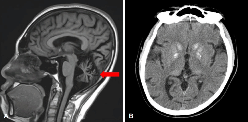
키는 150 cm (하위 10백분위수)였으며, 어머니에게 당뇨병과 난청이 있었고, 둘째 동생 역시 당뇨병이 있었다. 막냇동생은 발달장애 및 경련이 있었다. 임신 중에 혈당이 잘 조절되지 않았던 적이 있었으며, 8년 전 제1형 당뇨병으로 진단받아 인슐린을 사용하고 있었다. 6년 전 저혈당쇼크 이후 악화되어 매일 지속되는 두통, 어지럼, 구역, 구토, 기억력저하, 보행 실조, 방향감각저하로 본원 신경과에서 촬영한 뇌자기공명영상 및 CT상에서 소뇌위축 및 창백핵의 석회화 소견이 확인되었다(Fig. 3). 가족력이 있는 당뇨병, 난청, 소뇌실조 증상으로 미루어 미토콘드리아 질환을 의심하였고, 중합효소연쇄반응을 이용한 미토콘드리아 염기서열분석법으로 m.3243A→G 변이를 확인하여 MELAS 증후군으로 진단되었다.

MR and CT images. A: Sagittal MR T1 image show shows cerebellar atrophy which is indicated by red arrow. B: Axial CT image shows calcification on globus pallidus.
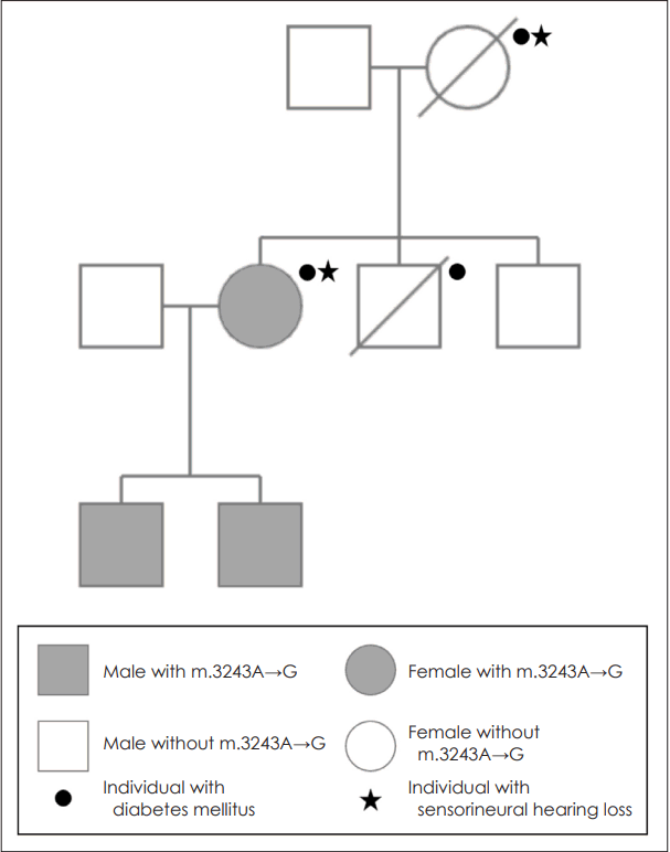
17세와 8세의 아들이 있으며, 17세의 아들은 저신장, 두통, 어지럼, 소뇌실조 증상이 있었고, 8세의 아들은 두통 어지럼 증상이 있었고, 중합연쇄반응을 이용한 미토콘드리아 염기서 열분석 검사를 통하여 두 명 모두 동일한 유전형의 MELAS 증후군으로 진단되었다(Fig. 4). 두 아들은 청력저하는 없었으나 첫째 아들의 뇌자기공명영상에서 경도 소뇌 위축 소견을 보였다. 본연구는 연구자 기관의 연구윤리심의위원회의 심의면제를 받았다.

Pedigree of patient. Small black circle represents those with diabetes mellitus and small black star represents those with sensorineural hearing loss. Family members diagnosed with mitochondrial encephalopathy, lactic acidosis, and stroke-like episodes (MELAS) syndrome was represented by gray filled shapes. Slashes denote deceased family members.
고 찰
MELAS 증후군은 미토콘드리아 DNA의 점 돌연변이에 의해 발생하며, 수정과정에서는 난자의 미토콘드리아만 전달되므로 모계유전의 특성을 가진다. 미토콘드리아 관련 질환은 환자마다 다양한 임상양상을 보이는데 이는 체세포 분열 시 변이된 미토콘드리아가 무작위적으로 분류되어 발생하는 이형질에 기인한다. MELAS 증후군은 미토콘드리아의 DNA 돌연변이에 의해 다양한 조직에서 미토콘드리아 호흡연쇄의 기능에 문제가 생겨 발생하는 것으로 알려져 있으며, 약 80% 에서 m.3243번째 염기의 점 돌연변이가 관찰된다[1]. 미토콘드리아 DNA 돌연변이와 관련된 질환의 증상은 에너지 요구량이 많은 기관인 와우, 중추신경계, 췌장, 골격근, 신세뇨관 등에서 발생하게 된다[7].
일본에서 보고된 연구에 따르면 3243A→G 변이의 빈도는 당뇨병 환자의 약 1%, 당뇨병과 난청을 같이 가진 환자에서 4.76%에서 발견되는 반면, 3243A→G 변이를 가지고 있는 당뇨병 환자의 61%에서 난청을 보인다[8]. MELAS 증후군에서 동반되는 난청은 후미로성 난청보다는 와우병변에 의한 난청으로 본 증례와 같이 청성뇌간반응검사상 무반응이더라도 어느 정도의 어음명료도를 보일 수 있는 이유가 된다[9]. 특히 와우에서 혈관줄무늬 세포는 높은 adenosine triphosphate 사용량을 요구하고, 분화하지 않기 때문에 미토콘드리아의 유전자변이에 따른 대사적 부담과 소음에 취약하다[9]. MELAS 증후군을 악화시키는 요인으로는 대사적 요구를 증가시키는 저혈당증, 신체적인 피로, 감염 등이 있을 수 있다[10]. 난청은 양측으로 진행하며, 점진적인 악화와 더불어, 계단식으로 대사성 스트레스를 유발하는 사건 이후 악화되는 경향을 보인다. 본 증례에서도 임신, 저혈당쇼크, 두부외상 등의 사건 이후로 난청의 발생 또는 악화가 되었다. 난청의 치료 방법으로 인공와우 이식이 있을 수 있는데, 한 연구에 따르면 미토콘드리아 병증으로 생긴 감각신경성 난청에서 인공와우의 이식으로 심도난청 환자 12예 중 7예에서 전화를 통한 소통이 가능하였으며, 나머지 5명도 이전보다 호전된 어음명료도를 보였다고 한다[11].
본 증례에서 시행한 비디오안진검사상 이상소견 보이지 않고 영상학적 검사상 소뇌위축이 확인되어 환자의 어지럼은 중추성으로 추정된다. 소뇌위축 및 운동실조는 MELAS 증후군에서 비교적 드물게 동반되는 비전형적 국내에서 소뇌위축이 동반된 MELAS 증후군 환자는 1예 보고된 바가 있다[6].
진행하는 양상의 양측성 감각신경성 난청이 당뇨병과 소뇌실조와 같은 신경학적 증상들을 동반하고, 대사성 스트레스의 상황에서 난청이나 신경학적 증상이 악화되는 양상을 보인다면 MELAS 증후군을 의심하고 미토콘드리아 염기서열 검사를 하는 것이 필요하다. 이형질에 의해서 환자들의 표현형 양상이 다양하기 때문에 미토콘드리아 질환에 대한 전반적인 이해와 표현형을 알고 가족력 등에 대해서도 파악하는 것이 진단에 중요하다. 또한 MELAS로 진단되었다면 소음과 감염, 대사성 스트레스를 예방할 수 있도록 교육하고, 지속적인 청력검사를 통하여 보청기나 인공와우 수술을 통해 청각재활을 도울 수 있다.
Acknowledgements
None
Notes
Author Contribution
Writing—original draft: Sun Seong Kang. Writing—review & editing: Woong-Woo Lee, Jung Ho Choi, Hyun Joon Shim.


