수포성 중비갑개의 세균구: 특이한 부위의 증례 보고
Bacterial Ball in Concha Bullosa: Report of a Case With Unusual Location
Article information
Trans Abstract
Choristoma, also known as a hairy polyp, is a rare benign mass that commonly occurs in the nasopharynx and oropharynx in the head and neck region. It is usually diagnosed in children and has rarely been reported in adults. In this study, we describe a nasopharyngeal choristoma in an adult man. The mass was located at the lateral nasopharyngeal wall, and the patient expressed intermittent nasal stuffiness and ear fullness. The mass was successfully removed using an endoscopic approach. Since nasopharyngeal choristoma in adults is rare, it is important to distinguish it from other benign tumors located in the nasopharynx or nasal cavity. In this report, we describe the radiologic characteristics of nasopharyngeal choristoma and summarize the importance of differential diagnosis from other benign masses.
Introduction
Bacterial balls are acellular mucous materials with bacterial colonies and inflammatory cell infiltrates, which were medically described in the paranasal sinuses (PNS) by Kim, et al. [1] in 2018. This rare disease was observed in the PNS of chronic rhinosinusitis (CRS) patients and is considered a new strategy for bacterial survival [1]. Since bacterial balls often involve a single paranasal sinus on one side and endoscopic findings during surgery are not characteristic, they are often confused with fungal balls [1,2]. Thus, their diagnosis can only be made using histopathological examination, in which multiple bacterial colonies and acellular materials are observed without fungi. In previous studies on bacterial balls, Kim, et al. [1] reported 6 cases in 2018 and Boiko [2] reported 20 cases in 2019, all occurring in the PNS.
Herein, we reported a case of a 47-year-old male with a concha bullosa bacterial ball, which had a decisive difference from previous cases, as the bacterial ball occurred in the middle turbinate concha bullosa rather than in the PNS. This was thought to be the first case reported worldwide, and thus we reported this unusual bacterial ball case, along with a literature review.
Case
A 47-year-old male presented with complaints of a 1-year history of left nasal obstruction and purulent nasal discharge. He had no notable medical conditions, such as allergies, asthma, immune deficiency diseases, cancers, or nasal traumas.
Using nasal endoscopic examination, a hard, bulging mass covered with smooth mucosa was found occupying the space between the nasal septum and left inferior turbinate, deviating the nasal septum to the right (Fig. 1). Aside from this, the patient’s hematological and biochemical parameters were normal.

Preoperative nasal endoscopic views showed a large mass-like lesion in the left nasal cavity. Right side (A) and left side (B) of the nasal cavity. S, nasal septum; T, left inferior turbinate.
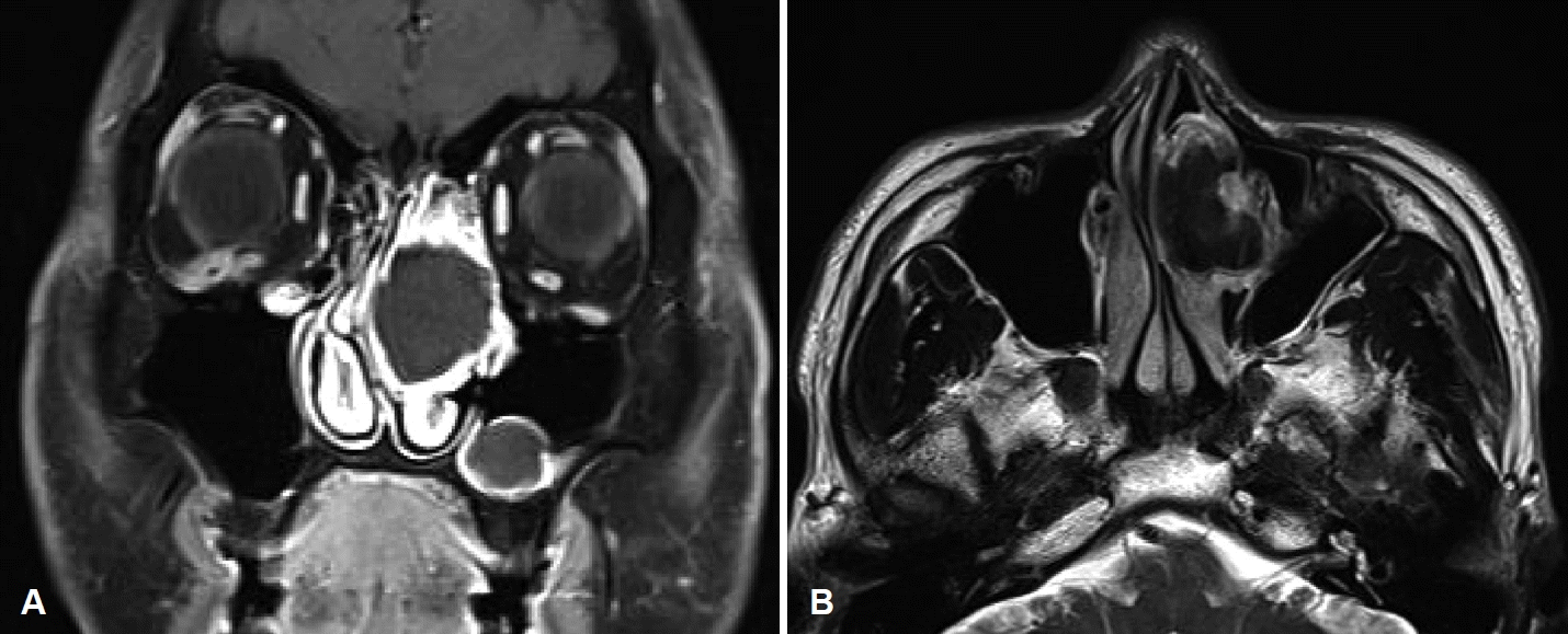
Contrast-enhanced CT of the PNS revealed a soft tissue density mass with peripheral rim enhancement and a central high-density lesion in the left nasal cavity, as well as a deviated nasal septum with adjacent bone remodeling (Fig. 2). On the other hand, MRI of the PNS revealed a well-defined mass displaying iso-signal features on T1-weighted images and obvious low-signal features on T2-weighted images, consistent with the findings of a fungal ball (Fig. 3).

Preoperative CT scan of paranasal sinuses. In the coronal view (A), a space-occupying lesion of soft tissue density was seen in the left nasal cavity with nasal septal deviation to the right side. In the axial and sagittal views (B and C), the lesion showed signs of expansile growth with adjacent bony remodeling. An internal high-density lesion was noted.

Preoperative MRI of paranasal sinuses. An enhanced T1-weighted image showed a lesion of iso-signal intensity with peripheral rim enhancement (A). A T2-weighted image demonstrated markedly low-signal intensity inside the lesion (B).
Under the impression of a left concha bullosa fungal ball with secondary left chronic maxillary sinusitis, the patient underwent functional endoscopic sinus surgery under general anesthesia. Using an endoscopic endonasal approach, an incision was made in the anterior part of the middle turbinate, the anterior wall was removed, and the left concha bullosa was found to be filled with green-colored semisolid materials (Fig. 4A and B). The materials were not easily broken and tightly adhered to the concha bullosa inner mucosa, making complete removal difficult. After carefully removing all of the materials, the involved middle turbinate mucosa was widely excised, allowing the infected materials and mucosa to be flushed from the nasal cavity (Fig. 4C).

Intraoperative nasal endoscopic views. Anterior surface of the mass-like lesion was incised (A) and the lesion was filled with greencolored, semisolid materials (B). The materials and involved middle turbinate were widely excised (C), and a white arrow indicates the anterior attachment of the middle turbinate. S, nasal septum; T, left inferior turbinate.
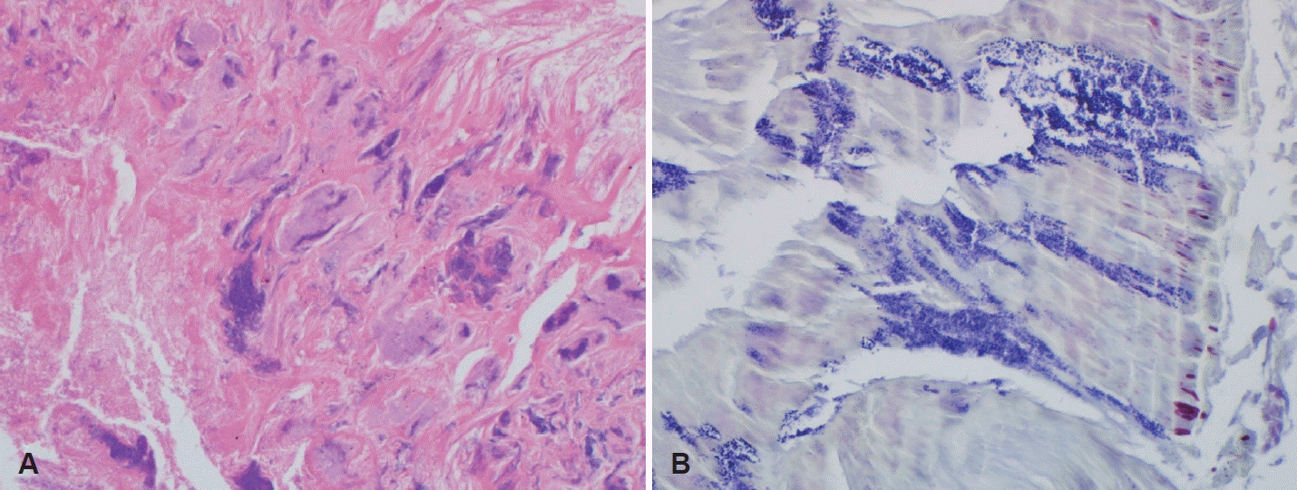
Contrary to our initial impression, no fungi were detected on microscopic, cultural, and histologic examinations. In hematoxylin and eosin (H&E) and Gram staining, multiple bacterial colonies and inflammatory cells were observed in the substrate composed of acellular mucous materials (Fig. 5), from which S. epidermidis was isolated in a biological culture test. The postoperative period was uneventful, and the patient was relieved from nasal symptoms. Furthermore, no signs of recurrence were noted during the 6-month postoperative nasal examination follow-up.
Discussion
Bacteria can form an organized microcolony surrounded by self-made extracellular polysaccharide matrix, which is defined as a biofilm. Bacterial biofilms have been though to play a major role in the pathogenesis of CRS, and can be detected using a confocal laser scanning microscopy or electron microscopy [3]. The concept of bacterial ball was first presented by Kim, et al. [4] in 2016; two unusual rhinosinusitis cases were found with macroscopic bacterial biofilms (bioballs) in the maxillary sinuses. In that study, the collected materials, which were suspected to be from maxillary sinus fungal balls, were found to consist of multiple bacterial colonies and an acellular matrix without fungi [4]. In 2018, Kim, et al. [1] also reported six cases of similar findings in the maxillary and ethmoid sinuses, suggesting that these materials be called bacterial balls. In contrast to a bacterial biofilm, which is observed under a microscope, a bacterial ball is a severe aggregate of bacterial colonies that can be observed macroscopically. Interestingly, the authors also suggested the possibility that biofilms can develop to be expressed as a bacterial ball, wherein the bacterial ball might be a new form of bacterial aggregation to increase survivability in the PNS [1,4].
Bacterial balls have been reported to account for 1.4% to 2.4% of CRS patients who underwent endoscopic sinus surgery in previous studies, clearly presenting a lower prevalence than bacterial biofilms or fungal balls [1,2]. In particular, CRS caused by bacterial balls is characterized by the appearance of single unilateral rhinosinusitis, commonly involving the maxillary sinus. As reported by Kim, et al. [1], 66% of bacterial balls occurred in the maxillary sinus and 33% in the ethmoid sinus, whereas in Boiko’s [2] study, bacterial balls occurred in 75% of maxillary sinuses and 25% of sphenoid sinuses. To our knowledge, since bacterial balls have been reported to occur in the paranasal sinus, this is the first report of a bacterial ball involved in the middle turbinate concha bullosa.
Bacterial ball diagnosis is made on the basis of CT scans, endoscopic findings in sinus surgeries, and biological and histopathological examinations, among which histopathology is the most important [1,2]. On PNS CT scans, bacterial balls are mainly located in the maxillary sinus of one side accompanied without central calcification in 75% of cases [1]. PNS MRI findings of bacterial balls have not been reported yet, but our study showed an iso-intense lesion in T1-weighted imaging and a low-signal lesion in T2-weighted imaging. Given that the imaging findings of bacterial balls are not specific as compared to those of fungal balls, it is difficult to differentiate between them prior to surgery. Thus, endoscopic sinus surgery is important for both diagnosis and treatment, wherein bacterial balls appear as gel-like or semi-solid materials with green or brown color. Additionally, bacterial balls were noted to be harder, less easily broken, and more tightly adherent to the mucosa of the PNS. Nevertheless, it is difficult to differentiate between the two groups at the time of surgery. Therefore, its definite diagnosis can only be made via histopathological examination, wherein bacterial balls show acellular mucous materials with bacterial colonies in H&E or Gram staining, without observable fungi findings [1,2]. In contrast, a diagnosis of fungal ball is based on histopathological findings with the presence of characteristic fungal hyphae without invasion of the mucosa on surgical materials [5].
Treatments for bacterial ball patients have not yet been established, but their complete removal from the infected mucosa is thought to be crucial, which is similarly done to treat fungal balls. Additional studies on the recurrence and treatment outcomes of bacterial balls are needed through continuous follow-ups.
The concha bullosa is a pneumatization and enlargement of a turbinate, occurring with an incidence of 14% to 54% in various studies [6]. This is usually asymptomatic and incidentally identified by CT scan; however, it can cause various symptoms, such as headache, pain between the eyes, or nasal obstruction, when it is extensively pneumatized. Moreover, it has a mucociliary transport system like other aerated paranasal cells draining into the frontal recess or middle meatus; thus, a problem with this drainage system leads to the development of various diseases in the concha bullosa [7,8]. Some of these differential diseases in the middle turbinate concha bullosa include mucoceles, mucopyoceles, fungal balls, cholesteatomas, and tumors [9]. Although bacterial balls in the concha bullosa have only been reported for the first time, they should be included in this list of differential diagnosis. Furthermore, entrapment and hypoventilation, which are caused by problems in the mucociliary transport system, appear to play an important role in bacterial ball development. Therefore, this should be considered when its pathophysiology is further investigated in future studies.
In conclusion, bacterial ball is a rare disease occurring in the PNS of CRS patients. Although bacterial balls in the concha bullosa are rare, they should be included in the differential diagnosis of patients with concha bullosa diseases.
Acknowledgements
None
Notes
Author Contribution
Conceptualization: Su-Jong Kim, Heung-Man Lee. Data curation: Jee Won Moon, Yongmin Cho. Investigation: Jee Won Moon. Methodology: Yongmin Cho. Supervision: Heung-Man Lee. Visualization: Jee Won Moon, Yongmin Cho. Writing—original draft: Su- Jong Kim, Heung-Man Lee. Writing—review & editing: Su-Jong Kim, Heung-Man Lee.

