양측 전두동의 공동 배출로: 증례 2예를 통한 이해
Common Drainage Pathway From Bilateral Frontal Sinuses: Lessons From 2 Cases
Article information

Trans Abstract
Primary pneumatization of the frontal bone occurs in the first year of life, but further pneumatization continues until 18 years of age. Elongation of the ethmoidal infundibulum and frontal recess, or upwards migration of the anterior ethmoidal cells has been proposed as a mechanism of frontal sinus development. The frontal sinus drainage pathway is displaced in the anterior or posterior direction depending on the pneumatization pattern of the frontoethmoidal cells. Also, frontal sinuses can be connected to the infundibulum or directly to the middle meatus depending on the superior attachment pattern of the uncinate process. Bilateral frontal sinuses are separated by their bony septum, and thus have their own drainage pathways. Contrary to this general rule, we experienced two cases of frontal sinuses that were connected to each other, showing a single drainage pathway from both frontal sinuses. We report these two cases along with a literature review.
서 론
전두동 배출로(frontal sinus drainage pathway)의 해부학적 구조는 복잡하기 때문에, 이비인후과 비부비동 내시경 수술 입문자에게는 도전해야 할 어려운 과제 중 하나이다. 비부비동 내시경 수술 중에 이러한 해부학적 구조와 변이에 대해 제대로 이해하고 해결하지 못하면 수술 후 부비동 폐쇄 및 의인성 전두동염의 원인이 될 수 있다[1,2]. 전두동 배출로 구조에 대한 체계적인 이해를 돕고자 2016년에 국제 전두동 해부 분류(International Frontal Sinus Anatomy Classification)가 출판되었으며, 해당 문헌에서는 전두동 배출로 주변의 전두사골봉소(frontoethmoidal cell)의 해부에 대한 체계적인 이해와 함께 수술 시 제거되는 구조물의 범위에 따른 전두동 수술법의 분류도 새롭게 제시되었다[3]. 저자들은 기존에 체계적으로 정리되고 알려진 일반적인 전두동 배출로 양상과 달리 양측 전두동이 연결되어 일측 비강으로 배출되는 증례들을 경험하였기에 문헌 고찰과 함께 보고하고자 한다.
증 례
증례 1
33세 남자가 인근 이비인후과의원에서 급성 부비동염 진단 하에 시행한 항생제 치료에 반응을 보이지 않아 의뢰되었다. 환자는 양측 코막힘, 화농성 비루와 후비루 증상을 호소하였으며 수양성 비루, 재채기, 코가려움 증상은 없었다. 기저질환 및 과거의 비강, 부비동의 수술 혹은 외상의 병력은 없었다. 비강 내시경 검사에서 좌측 중비도에 유두(papillary) 모양의 종물이 발견되었다. 부비동 컴퓨터단층촬영(paranasal sinus computed tomography, PNS CT)에서 양측 전두동과 사골동 및 상악동 내에 연조직 음영이 확인되었으며, 양측 전두동이 중격의 결손을 통해 연결되어 있었다(Fig. 1A and B). 좌측 전두동은 비제봉소(agger nasi cell)와 사골포(ethmoid bulla) 사이를 통해 사골누두(ethmoidal infundibulum)로 통하는 배출로가 관찰되었으나(Fig. 1C) 우측 전두동은 우측 중비도 혹은 사골누두로 통하는 배출로를 확인할 수 없었다(Figs. 1D and 2). 우측 만성 비부비동염 및 좌측의 반전성 유두종을 동반한 만성 비부비동염 진단하에 부비동내시경 수술을 시행하였다. 우측 사골동절제술 및 중비도상악동개방술 시행 후 전두와(frontal recess) 부위에서 전두동 배출로가 존재하지 않음을 내비게이션(navigation)을 이용하여 재확인하였으며, 좌측의 경우 구상돌기(uncinate process) 기원의 종양을 확인한 후 정상 부비동 점막과의 충분한 절제연을 두고 종양을 완전히 절제하였고, 사골동절제술 시행 후 전두동 배출로를 확인한 뒤 넓게 개방해 주었다. 수술 후 2개월째 환자는 특별한 증상을 호소하지 않았으나 비강내시경 검사 시 좌측 전두동 배출로 부위의 점막 부종 소견이 있었으며(Fig. 1E), Waters 영상에서 우측 전두동의 혼탁 소견이 관찰되었다(Fig. 1F). 수술 후 3개월째 추적 관찰 시 비강 내시경 검사에서 좌측 전두동 및 배출로는 정상 점막 소견을 보였고(Fig. 1G), Waters 영상에서도 양측 전두동 모두 정상 소견을 보이는 것을 확인하였다(Fig. 1H).
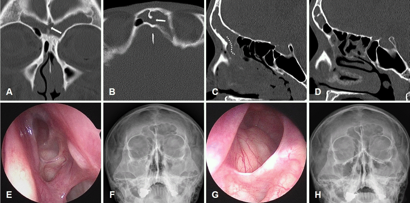
Representative images of case 1. A and B: In CT scans, both frontal sinuses, which were filled with soft tissue densities, were connected through a bony defect between them (arrows). C: The left frontal sinus drainage pathway (dotted line) was observed between the ager nasi cell and ethmoid bulla. D: In contrast, the right frontal sinus drainage pathway was not identified, which was confirmed by the help of the navigation system during surgery. E: At postoperative two months, in the endoscopic view, the left ethmoid cavities were clear, but the frontal sinus drainage pathway was covered by swollen mucosa. F: In the Water’s view, the left frontal sinus appeared clear, but the right frontal sinus showed haziness. G and H: At postoperative three months, the left frontal sinus was clear and the drainage pathway was patent (G), and the right frontal sinus turned clear in the Water’s view (H).
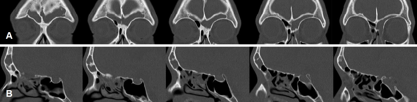
Serial coronal (A) and right sagittal (B) CT images of case 1. The right frontal sinus drainage pathway to the right ethmoid sinus is not seen.
증례 2
28세 남자가 발치 후 수개월간 지속된 우측 코막힘을 주소로 인근 이비인후과의원에서 2개월간 약물 치료를 받았으나 호전이 없어 내원하였다. 수양성 비루, 재채기, 코가려움 증상은 없었으며, 기저질환 및 과거의 비강, 부비동의 수술 혹은 외상의 병력은 없었다. 비강 내시경 검사에서 우측 중비도의 화농성 비루가 관찰되었다. PNS CT에서 우측 전두동과 사골동 및 상악동 내의 연조직 음영이 확인되었으며, 양측 전두동은 중격 결손을 통해 연결되어 있었다(Fig. 3A and B). 우측 전두동에서는 비제봉소와 사골포 사이를 통해 사골누구로 통하는 배출로가 확인되었으나(Fig. 3C), 좌측 전두동에서는 좌측 중비도 혹은 사골누두로 통하는 배출로를 확인할 수 없었다(Figs. 3D and 4). 아목시실린/클라불란산(ratio of 7:1)과 3세대 세팔로스포린 계열 항생제를 각각 2주간 사용하였으나 증상이 전혀 호전되지 않아 우측 치성 부비동염 진단하에 부비동 내시경 수술을 시행하였다. 우측 사골동 절제술 후 전두동 배출로를 확인한 후 넓게 개방해 주었으며, 좌측 전두동의 배출로가 존재하지 않음을 내비게이션을 이용하여 재확인하였다. 수술 후 2개월째 환자의 주관적인 증상은 호전 중이었으나 비강 내시경 검사 시 우측 전두동 배출로 부위의 점막 부종 소견이 있었으며(Fig. 3E), Waters 영상에서 양측 전두동의 혼탁 소견이 관찰되었다(Fig. 3F). 수술 후 3개월째 추적 관찰 시 비강 내시경 검사에서 우측 전두동 및 배출로는 정상 점막 소견을 보였고(Fig. 3G), Waters 영상에서도 양측 전두동 모두 정상 소견을 보였다(Fig. 3H).
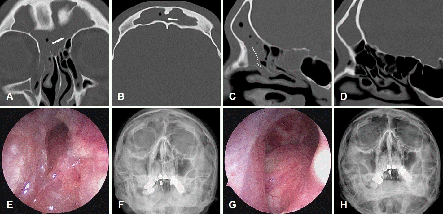
Representative images of case 2. A and B: In CT scans, both frontal sinuses, which were filled with soft tissue densities, were connected through a bony defect between them (arrows). C: The right frontal sinus drainage pathway (dotted line) was observed between the ager nasi cell and ethmoid bulla. D: In contrast, the left frontal sinus drainage pathway was not identified, which were confirmed by the help of the navigation system during surgery. E: At postoperative two months, in the endoscopic view, the right frontal sinus drainage pathway was covered by swollen mucosa. F: In the Water’s view, both frontal sinuses showed haziness. G and H: At postoperative three months, the right frontal sinus was clear and the drainage pathway was patent (G), and both frontal sinuses turned clear in the Water’s view (H).
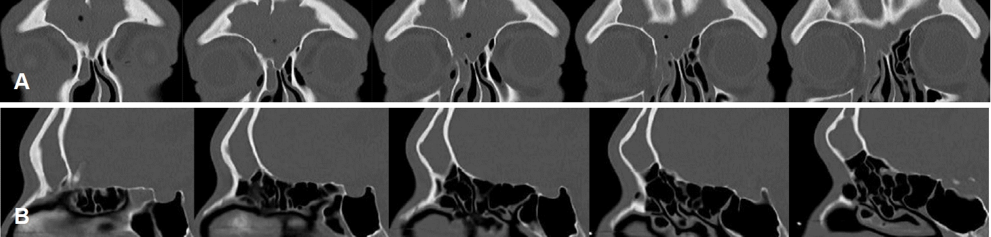
Serial coronal (A) and left sagittal (B) CT images of case 2. The left frontal sinus drainage pathway to the left ethmoid sinus is not seen.
고 찰
전두동은 전두골이 함기화된 것으로 전두와 전체가 전두골 방향으로 확장되어 생성되거나, 발생 당시 전두와에 있던 4개의 와(pit) 중 하나에서부터 생성된다. 그리고 중비도의 사골누두가 상부로 확장되어 생성되거나, 또는 사골포로부터 함기화됨으로써 발생한다[4]. 한편, 전두동의 전내측으로부터 자연 배출로를 통해 중비도로 이행되는 부위는 관이라기보다는 상하의 깔때기 모양이 만나 좁아지는 구멍 형태로 이루어져 있다. 전두동으로부터 중비도로 배출되는 경로는 구상돌기와 사골포 기판의 형태 및 부착부위에 따라 전두와, 사골누두 등으로 다양하다[5]. 또한 전두동 배출로는 상비제봉소, 상비제전두봉소(supra-agger frontal cell)의 발달에 의해 후방으로 전위되거나 상사골포봉소(supra-bullar cell), 상사골포전두봉소(supra-bullar frontal cell)의 발달에 의해 전방으로 전위되기도 한다[6]. 하지만 저자들이 광범위한 문헌 검색을 했음에도 불구하고 양측 전두동이 공동의 일측 배출로를 갖는 경우는 기존에 보고된 바가 없었다.
본 연구의 증례 1에서는 우측 사골누두나 중비도로 연결되는 우측의 전두동 배출로를 확인할 수 없었으며, 양측 전두동 사이 중격의 결손을 통해 양측 전두동 모두 좌측 전두동 배출로를 통해 배액이 되는 구조임을 추정할 수 있었다. 이러한 구조에서는 양측 전두동이 일측으로 배액될 것이라는 가설을 세울 수 있었으며 좌측의 전두동 개방술만 시행했음에도 불구하고 수술 후에 환자의 증상과 Waters 영상에서 양측 전두동의 혼탁 소견이 모두 소실된 것이 이러한 가설이 옳았음을 증명한다. 증례 2의 경우 우측 상악동의 치성 부비동염이 우측 사골동과 전두동으로 파급이 되었으며 전두동 중격의 결손을 통해 좌측 전두동으로 파급되었던 증례로 생각되며, 좌측의 사골동, 상악동, 접형동 모두 PNS CT에서 정상 소견을 보였다(Fig. 3A and D). 증례 1과 마찬가지로 일측의 전두동 개방술을 통해 양측 전두동의 병변이 해소될 수 있으리란 가설을 세울 수 있었으며, 우측 전두동 개방술만으로도 수술 후 환자의 증상과 Waters 영상에서 양측 전두동의 혼탁 소견이 모두 소실된 것을 통해 이러한 가설이 옳았음을 확인할 수 있었다.
상기의 두 증례 모두에서 전두동 중격의 결손이 발생할 만한 외상 혹은 수술의 병력은 없었으며 동측의 배액로가 없는 전두동의 점액섬모운동이 반대측 전두동을 향했기 때문에 별도의 전두동 개방술 없이 병변이 호전되었을 것으로 생각된다. 따라서 두 증례 모두 지속적인 염증 혹은 다른 원인에 의한 2차적인 골결손보다는 전두동의 발달 과정에서 전두동 중격 결손이 일어났을 것으로 추정된다. 전술한 바와 같이 양측 전두동이 공동의 일측 배출로를 갖는 증례는 이전에 보고된 바가 없었지만, 저자들은 추가적인 문헌 검색을 통해 일측 전두동의 무형성(aplasia)이 반대측 전두동의 과함기화로 인해 드러나지 않는 숨은 무형성(hidden aplasia) 증례에 대한 영상학적 연구를 확인할 수 있었다[7]. 즉, 일측의 숨은 전두동 무형성과 반대측 전두동의 과함기가 마치 중격에 결손을 동반한 양측 전두동처럼 보이는 사례가 있다는 것이며, 저자들이 경험한 2예도 이에 해당하는 것으로 볼 수 있다. 해당 연구에서는 305예의 CT 영상을 분석하여 3.6%에서 일측 전두동의 숨은 무형성을 확인할 수 있다고 보고하였다. 저자들은 이와 같은 증례를 지난 10여년간 3300예 이상의 부비동 내시경 수술 중 단 2예에서만 경험하였는데, 이러한 발생률의 차이는 영상학적 검사에서는 일측의 전두동 무형성이 드물지 않지만 내시경 전두동 수술을 요하는 병변을 동반하는 환자는 그 중 극히 일부에 지나지 않는다는 점을 시사한다. 따라서, 비록 실제 임상에서 문제가 되는 경우는 드물겠지만 이러한 해부학적 변이에 대해 인지하지 못함으로써 불필요한 건측의 사골동 수술을 시행하거나 숨은 전두동 무형성이 있는 비강에서 무리한 전두동 개방술을 시도하다 두개저 손상과 같은 합병증을 초래할 가능성이 있으므로 일측 전두동 무형성이라는 해부학적 변이에 대해 인지할 필요가 있겠다. 한편, 비중격만곡증 등을 비롯한 여러 해부학적 변이 중 일측 전두동 무형성과 연관된 위험인자는 알려진 바가 없으며 저자들이 경험한 두 증례에서도 양측 비강 및 부비동의 부피의 차이 혹은 비대칭 등 뚜렷한 해부학적 차이는 확인할 수 없었다. 추후 보다 많은 환자들을 대상으로 한 CT 영상 분석과 사체 연구 등을 통해 일측의 공통 배출로를 갖는 양측 전두동에 대한 보다 체계적인 연구가 필요할 것으로 생각된다.
Acknowledgements
This work was supported by the grant of Institute of Health Sciences of Gyeongsang National University (HIS GNU-2020-03).
Notes
Author Contribution
Conceptualization: Ki Ju Cho, Sang-Wook Kim. Data curation: Ki Ju Cho, Sang-Wook Kim, Sang Yun Lee. Funding acquisition: Sang-Wook Kim. Investigation: Sang Yun Lee, Ki Ju Cho. Supervision: Sang-Wook Kim. Validation: Yung Jin Jeon, Sang-Wook Kim. Writing—original draft: Ki Ju Cho, Sang Yun Lee. Writing—review & editing: Sang-Wook Kim.