74세 남자 환자에서 비순 낭종으로 오인된 비강저에서 발생한 혈관성 평활근종
Vascular Leiomyoma Arising From the Nasal Floor Misdiagnosed as a Nasolabial Cyst in an 74-Year-Old Patient
Article information
Trans Abstract
Leiomyoma is a benign tumor that originates from smooth muscle and can develop anywhere in the body where smooth muscle is present. However, leiomyoma arising in the head and neck region are very rare. We experienced a case of a 74-year-old male patient with a right nasal floor mass. The patient complained of bleeding from the right nasal cavity, nasal congestion, and discharge. On physical examination, a relatively smooth-surfaced, well-demarcated mass was observed in the right nasal floor. Preoperative imaging was interpreted as a right nasolabial cyst, and endoscopic excision was performed. Histologic examination confirmed it to be vascular leiomyoma. The patient was followed up for 6 months postoperatively and remains stable with no recurrence of symptoms. We report a case of vascular leiomyoma arising from the nasal floor that was mistaken for a nasolabial cyst in a 74-year-old patient.
서 론
평활근종은 평활근에서 기원하는 양성종양으로 신체에서 평활근이 존재하는 곳이라면 어느 곳에서나 발병할 수 있다고 알려져 있다. 그중에서도 자궁을 비롯한 여성 생식기계에서 전체 증례 중 약 95% 정도의 빈도로 흔하게 발병하며, 그외에는 피부, 위장관계에서 주로 발생하는 것으로 알려져 있다[1]. 두경부 영역에서 발생하는 평활근종은 전체 평활근종 중 1% 미만이며, 그중에서도 비강 및 부비동에서는 3% 미만의 빈도로 매우 드문 종양으로 알려져 있다[2]. 평활근종은 비혈관성 평활근종(leiomyoma), 혈관성 평활근종(angioleiomyoma), 그리고 상피양 평활근종(epitheloid leiomyoma)의 3가지 형태로 나누어지며, 현재까지 보고된 바에 따르면 비강 내에서 발견된 평활근종은 대부분 혈관성 평활 근종(86.1%)으로 보고되었다[3]. 저자들은 비순낭종으로 오인되었던 비강저(nasal floor)에서 발생한 혈관성 평활근종 1예를 경험하였기에 문헌 고찰과 함께 보고하고자 한다.
증 례
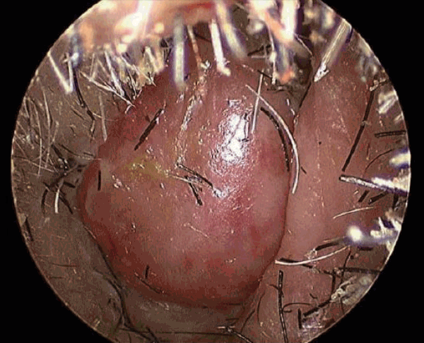
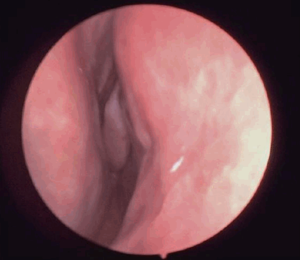
74세의 남자 환자가 내원 수개월 전부터 발생한 우측 비강의 출혈 및 코막힘을 주소로 내원하였다. 출혈은 매일 간헐적으로 발생하였고 심한 경우에는 노란 분비물도 동반되는 증상이 있었다. 과거력상 고혈압, 뇌경색, 심방세동이 있었으며, 이로인한 우측 반신마비 병력이 있었으며 항혈소판제를 복용 중이었다. 비강 내시경 검사상 우측 비강 저부에 존재하는 종물이 관찰되었다(Fig. 1). 그 외 비인두, 구강, 후두 및 경부 등 다른 신체검사상에서는 별다른 특이소견은 보이지 않았다. 종물은 표면이 매끄럽고 비교적 경계가 명확한 구형의 형태로 관찰되었으며, 우측 비강바닥 부위에 부착되어 있는 양상이었다. 종물의 크기 및 파급 범위에 대한 추가적인 평가를 위해 부비동의 조영 전산화단층촬영검사를 시행하였다. 검사 결과 우측 비강저 부위에 전체적으로 저음영을 띠고 있으며 약간의 주변부 조영증강이 관찰되는 1.5×2.3×2.2 cm 크기의 종물이 확인되었다(Fig. 2). 종물은 우측 비강 하부를 거의 막고있는 양상으로 비중격의 우측 편위가 동반되었다. 신체검진 및 영상 검사 소견으로 미루어 볼 때 비순낭종으로 진단 후 수술적 제거를 계획하였다. 먼저 내시경을 이용하여 종물을 확인한 후 종물의 기시부를 포함하여 종물을 완전히 절제하였다. 이후 흡입 코블레이터를 이용하여 지혈을 시행하였다. 종물은 출혈이 많지 않고 주변 조직과 유착이 심하지 않아 비교적 쉽게 제거되었으며, 종물을 덮고 있던 점막의 손상도 없었다. 제거된 병변은 단단하고 둥근 형태로 회백색을 띠고 있었다(Fig. 3).
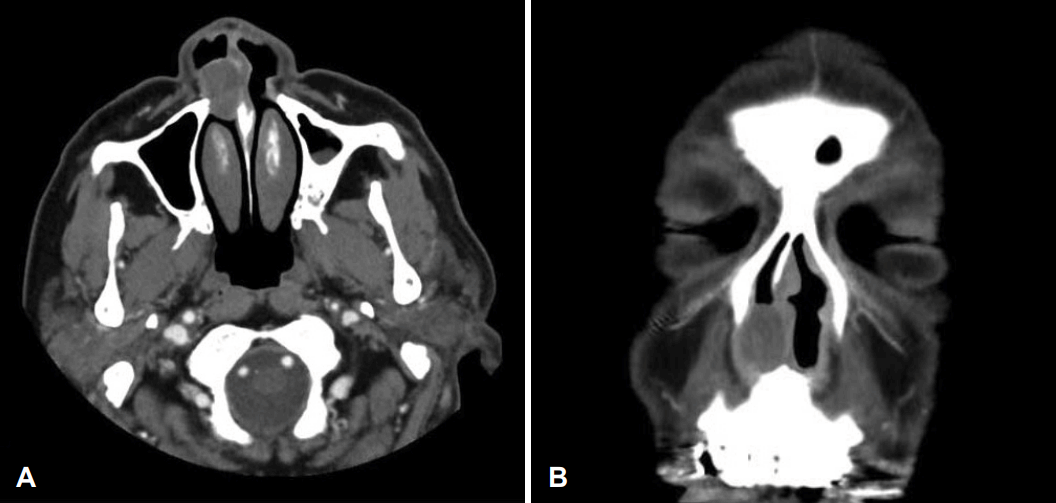
Enhanced paranasal sinus CT shows a mass originating at the right nasal floor that measures 1.5×2.3×2.2 cm in size. A: Axial view. B: Coronal view.
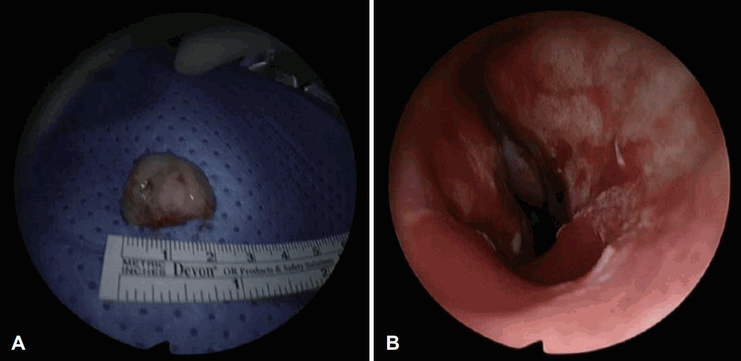
Intraoperative endoscopic images. A: Gross specimen was completely removed after surgery. B: Nasal endoscopy shows the Rt nasal cavity where tumor has been removed.
병리 소견상 다양한 방추형 세포들이 서로 교차하여 다발을 이루고 있으며 일부에서 혈관벽이 관찰되었다(Fig. 4A). 면역 조직 화학 염색상 종양세포는 민무늬근육 액틴(smooth muscle actin)에 강양성 반응을 보였다(Fig. 4B). 이러한 병리 소견을 종합하여 최종적으로 혈관성 평활근종으로 진단되었다. 환자는 수술 후 6개월째 추적 관찰 중이며, 주관적 증상은 없고 비내시경 검사상에서도 재발 소견도 보이지 않고 있다(Fig. 5).
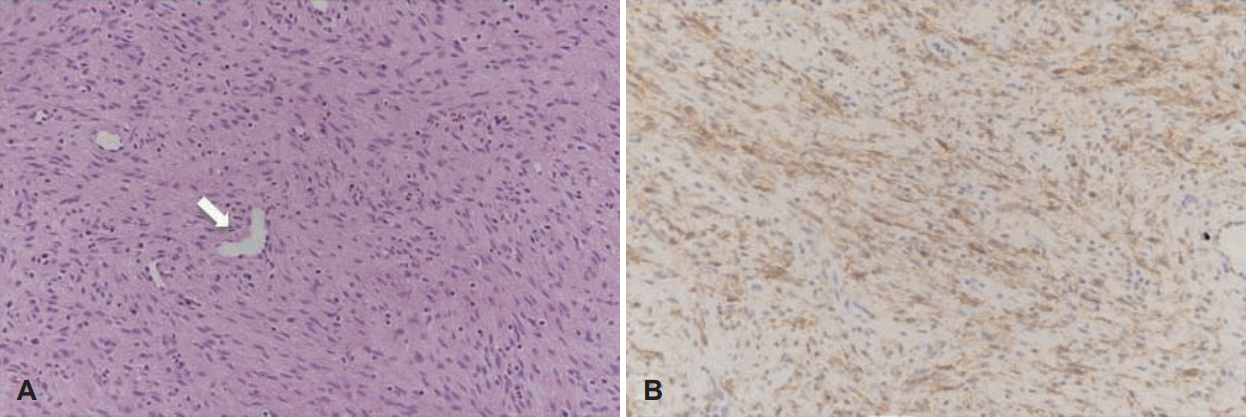
Histopathologic findings of right nasal cavity mass. A: Immunohistochemistry demonstrates that the mass was composed of spindle cells with irregularly dilated, staghorn-shaped vessels (arrow) (H&E stain, ×100). B: The tumor cells were positively stained for smooth muscle actin (brown color, ×100).
고 찰
두경부영역에서 혈관성 평활근종은 매우 드물게 보고되어 왔다. 그중에서도 부비강 부위는 더욱 드문 발병 위치이다[4]. 비강 내부에서 발생한 혈관성 평활근종에 대한 보고는 비교적 다수 있었으나, 비강저에서 발생한 혈관성 평활근종의 경우 현재까지 국내에서 1예만이 발표되었다[5].
평활근이 거의 존재하지 않는 비강에 평활근종이 발생하는 원인에 대해서는 아직까지 명확히 밝혀져 있지 않다. 비강 내 미분화 중간엽(aberrant undifferentiated mesenchyme)에서 기원하였다는 가설, 혈관벽의 평활근에서 발생하였다는 가설, 마지막으로 비전정의 기모근(elector pilae muscle) 혹은 땀샘에서 기원하였다는 가설이 존재한다[3,6,7].
비강 및 부비동에서 발생하는 평활근종은 성장속도가 느리기 때문에 일정 크기 이상 증가하기 전까지는 비특이적인 임상 양상을 보이며, 종양의 위치와 크기에 따라 나타나는 증상이 다르다. 이 때문에 두경부 영역에서 발생한 혈관성 평활근종의 경우 30%에서 무증상을 보이는 것으로 보고되었다[2]. 이러한 평활근종이 유발하는 증상으로는 주로 비폐색, 종양 부위 동통, 두통, 비건조감, 비출혈 등을 초래할 수 있다고 알려져 있다[8,9].
평활근종은 전산화단층촬영상에서 약한 조영증강을 보이며 비교적 균질한 양상의 종물로 관찰되며, 자기공명영상검사상에서는 조영증강을 보인다고 알려져 있다[2]. 그러나 평활근종을 진단할 만한 특징적인 소견이 없어 영상검사가 확진에 유용하지 않으며, 세침흡인 검사와 세포 검사 역시 평활근종을 확진할 수 없다[2]. 이러한 임상적 특징으로 인해 수술을 통한 조직학적 확진 이전에 부비동 내의 혈관성 평활근종을 진단하는 것은 어렵다[10]. 평활근종은 혈관종(hemangioma), 혈관섬유종(angiofibroma) 및 신경성 종양과 같은 간엽 기원의 종양과의 감별이 필요하며[3] 이외에도 다형성 선종(pleomorphic adnoma), 비순낭종 등과 같은 기타 양성종양, 그리고 섬유육종(fibrosarcoma), 평활근육종(leimyosarcoma) 등과 같은 연조직 악성종양 등과 같은 질환과 감별하여야 한다[11]. 조직병리학적 검사가 혈관성 평활근종을 진단할 수 있는 가장 정확한 검사이다. 혈관성 평활근종의 조직학적 소견으로 hematoxylin and eosin 염색상에서 길쭉한 내부에 끝이 뭉툭한 핵을 가지는 방추형 모양을 이루는 평활근 세포의 다발과 함께 두꺼운 혈관벽이 관찰된다[2,12]. 혈관종, 혈관섬유종과 같은 다른 방추형 세포 종양과의 감별을 위해서 평활근 세포(actin, desmin)와 혈관 내피세포(CD31, CD34, factor VIII)에 대한 면역조직화학 염색이 이용될 수 있다[2].
혈관성 평활근종의 치료는 완전한 수술적 적출술이 원칙이다[10]. 기존 연구에서는 적출술, 내시경적 절제술, Caldwellluc 수술 등이 제안되었다. 종양의 크기 위치 및 범위에 따라 수술 방법이 결정되며, 종양이 비강에 국한된 경우 본 예와 같이 비내시경을 이용한 제거가 선호된다. 수술 후 악성 변화나 재발은 보고된 바가 없다[2].
본 증례에서는 수술 전 영상검사 및 신체 검진상 비순 낭종으로 진단된 상태로 수술을 진행하였으며, 술후 진행한 조직 검사상에서 평활근종을 확진할 수 있었다. 평활근종의 경우 비강내에서 발생하는 경우는 매우 드물 뿐만 아니라 그중에서도 비강 바닥에서 발생하는 사례는 더욱 드물다고 알려져 있다. 뿐만 아니라 영상학적으로 특징적인 소견이 없어 수술 전 진단이 어렵다. 그렇기 때문에 본 임상 사례와 같이 다른 종양으로 오인하여 수술한 이후 조직검사에서 확진이 되는 경우가 많다. 따라서 비강 내 종물을 주소로 내원 한 환자를 진료할 때 이점을 유념해야 할 것으로 생각되며, 그 의의가 있다고 생각하여 보고하는 바이다. 환자는 수술 후 6개월동안 추적 관찰 중이며 현재까지 재발의 징후 등 특이소견은 보이지 않았다.
Acknowledgements
None
Notes
Author contributions
Conceptualization: Kun Hee Lee. Data curation: Jong Hwan Lee, Seung Yup Son. Formal analysis: Jong Hwan Lee. Methodology: Kun Hee Lee. Project administration: Seung Yup Son. Supervision: Kun Hee Lee. Writing—original draft: Jong Hwan Lee. Writing—review & editing: Kun Hee Lee, Jong Hwan Lee.

