추체 첨부 거대 선천성 진주종의 경미로 조대술 치험 1예
A Case of Huge Congenital Cholesteatoma of Petrous Apex Treated With Translabyrinthine Marsupialization
Article information

Trans Abstract
Congenital cholesteatoma is rare, and especially so for a benign mass to grow from the squamous epithelium and the petrous apex as a location for this cancer. Surgery of the petrous apex cholesteatoma is challenging due to the location of the lesion and the need to preserve adjacent structures such as the dura mater, internal carotid artery, and internal auditory canal. Imaging studies, including CT and MRI, were crucial in guiding the surgical approach. This report describes a 40-year-old male patient with a large congenital cholesteatoma involving the petrous apex and temporal bone. The patient presented with a headache, hearing loss that persisted despite prior medical treatment. The cholesteatoma was surgically marsupialized through translabyrinthine approach, and the patient has remained stable for over 20 years. This report underscores the importance of considering the anatomical location and relationships of the lesion and adjacent structures when determining the surgical approach for congenital cholesteatoma involving the petrous apex and temporal bone.
서 론
선천성 진주종은 전체 진주종 중 2%-5%를 차지하며 비교적 드문 질환이다[1]. 그중에서도 추체 첨부는 측두골의 내측 부분으로 접형골과 후두골 사이에 있는 곳을 말하며 진주종 발생이 특히 드문 곳으로 알려져 있다[2].
선천성 진주종의 치료는 모든 병변을 제거하여 염증과 합병증 위험이 없는 귀를 만들고 재발이 예방되어야 하며 청력의 보존을 목표로 한다[3]. 하지만 추체골은 경막, 내이도, 내경동맥 등과 인접해 수술적 접근방식에 제한과 어려움이 있고, 주위 구조물을 보존하며 병변을 모두 제거하는 것을 어렵게 한다[4]. 따라서 병변의 해부학적 위치, 중요 구조물과의 관계를 고려하여 수술 방법과 치료의 방향을 결정하여야 한다[5].
본 증례에서는 40세 남환에서 발생한 추체 첨부와 추체골을 침범한 거대 선천성 진주종에 대해 경미로 접근법을 이용한 조대술을 통해 치료 후 20년의 장기간 추적 관찰 기간동안 안정적으로 유지되고 있는 1예에 대해 문헌 고찰과 함께 보고하는 바이다.
증 례
40세 남자 환자가 10년 전부터 발생한 좌측 난청과 두통으로 내원하였다. 환자는 30대부터 상기 증상이 있었으나 특별한 검사 및 치료 없이 지내왔다. 환자는 두통에 대한 1차 병원 치료에도 호전되지 않았다. 좌측 난청과 이명이 동반되었으나 어지럼증은 호소하지 않았다. 환자는 0.5갑씩 20년의 흡연력이 있으며 1990년 결핵 치료받은 과거력 이외에 귀 수술 병력은 없었다.
이경 검사상 좌측 고막에 중등도의 중심부 천공과 함께 이루와 육아조직이 관찰되었다(Fig. 1). 좌측 이루에서 시행한 균 검사상 메티실린 내성 황색포도알균이 동정되었다.
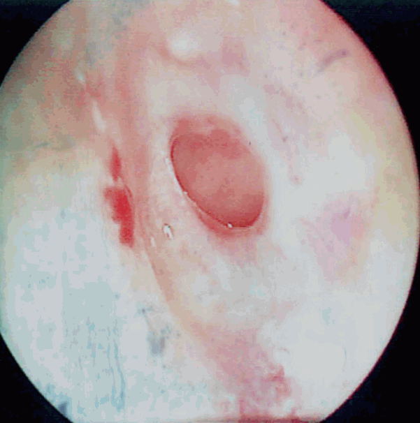
Left tympanic membrane. Serous discharge and moderate central perforation is noted.
순음 청력 검사 결과상 좌측 기도 청력 역치 105 dB, 골도 청력 역치 59 dB로 좌측에 심도의 혼합성 난청 소견이 관찰되었다(Fig. 2).
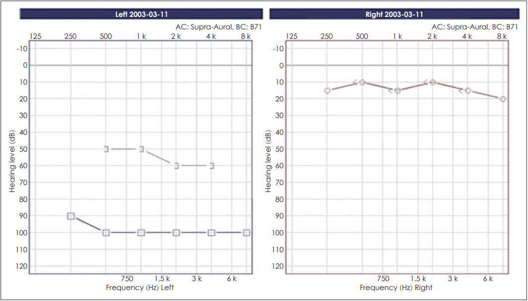
Pure tone audiometry shows profound hearing loss in the left ear with a pure tone threshold 105 dB.
측두골 전산화단층촬영 검사에서 좌측 추체골을 점유하고 있는 2.7×2 cm의 연부조직 음영이 관찰되었고 유양돌기동에서부터 고실까지 연부조직 음영이 가득 채우고 있었으며 이소골과 내이의 침윤 및 미란도 관찰되었고(Fig. 3), 자기공명영상촬영에서는 병변 부위가 T2 강조영상에서는 고강도 신호, 조영증강 하지 않은 T1 강조영상에서는 중등도의 신호가 관찰되며 gadolinium 조영증강 T1 강조영상도 중등도의 신호가 관찰되어(Fig. 4), 이낭을 포함하고 추체 첨부부터 미로의 상하부를 모두 포함하는 Sanna Class IV에 해당하는 선천성 추체골 진주종 소견을 보였다[6].

Computed tomographic images of left temporal bone showing soft tissue density on petrous apex and mastoid and middle ear cavity. The lesion eroded labyrinth and inner ear structure and encase petrous part of internal carotid artery axial image (A), coronal image (B).
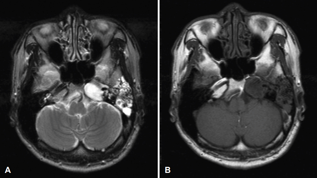
MRI scan demonstrating lesion of the left petrous apex. T2-weighted axial MRI scan (A), T1-weighted axial MRI scan (B).
추체 첨부에 발생한 선천성 진주종에 의해 내경 동맥 등 중요 구조물들이 노출되어 있어 진주종의 완전한 제거는 어렵다고 판단하여 경미로 접근법을 이용하여 조대술을 계획하였다.
전신마취하 경미로 접근법을 이용한 광범위 유양돌기 삭개술 및 추체골절제술을 통해 진주종의 근전절제와 조대술을 시행하였다. 수술 당시 중이는 육아 조직으로 채워져 있었으며 진주종에 의해 파괴된 이소골과 구조물은 제거하였으며 진주종으로 인한 경막의 손상은 관찰되지 않았다. 외측 반고리관과 난원낭을 제거 후 추체 꼭지 내부로 이어지는 거대한 진주종낭이 있었으며 진주종을 충분히 제거 후 진주종낭의 입구를 유지하기 위해 근막 이식물을 삽입하였다. 달팽이관이 소실된 부위는 이개 연골을 이용하여 채우고 안면 신경도 우측 둔부의 피부로부터 부분층피부이식술을 이용하여 덮어 주었다.
수술 후 병리 결과로 육아조직과 함께 진주종이 확인되어 선천성 진주종을 진단할 수 있었다.
술후 두통은 호전되었으며 전신상태 양호하였고 안면마비 및 뇌수막 이탈, 뇌척수액 누출, 뇌농양 등의 합병증은 관찰되지 않아 퇴원 후 외래 추적 관찰하였다. 술후 5년 1개월 후 시행한 측두골 전산화단층촬영 검사에서 병소 양호한 소견을 보였다(Fig. 5).

Postoperative axial image (A) and coronal image (B) of CT shows well marsupialized cavity after 5 years.
수술 후 6년 6개월째 경미로 접근법을 시행한 부위에 육아조직으로 인해 협착이 발생하여 내부 염증 의심되어 진주종낭 입구에 실라스틱 배액관을 삽입하였고 외래에서 주기적으로 교체하며 병소 양호한 것을 확인하였다.
이후 주기적으로 외래에서 실라스틱 배액관을 교체하며 추적 관찰하였으며 수술 후 20년 후 시행한 전산화단층촬영과 내시경상 진주종 재발없이 병소 안정적으로 유지되고 있다(Fig. 6).
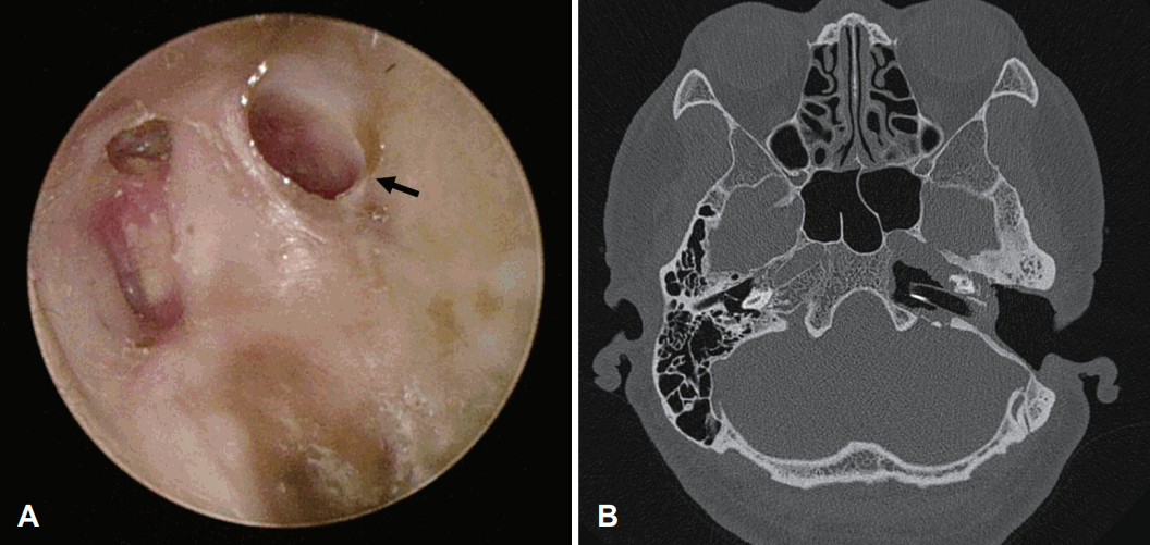
Postoperative otoscopic findings of left tympanic membrane. A: The translabyrinthine approach tract (black arrow) is noted. B: Postoperative axial image of CT shows well marsupialized cavity after 20 years.
고 찰
측두골에 발생하는 선천성 진주종은 원발하는 부위에 따라 중이, 고막, 외이도, 유앙돌기 또는 추체부 진주종으로 분류할 수 있다[3]. 그중에서도 선천성 추체부 진주종은 매우 드물며 전체 진주종 중 1%-3%를 차지하고 있고 발생기전은 태생기 외배엽 유래의 상피세포가 남아 추체부로 이동하여 각질화 상피세포로 증식하여 발생한다고 알려져 있다[3].
진주종의 치료는 외과적 절제술이 원칙이며 병변의 위치와 범위를 참고하여 실험적 고실 개방술, 고실 성형술, 또는 유양돌기 절제술 등을 적절히 선택해야 한다[7].
측두골 추체부 진주종은 두부의 아주 깊은 심부에 위치하기 때문에 오랜 기간 증상을 나타내지 않다가 진주종에 의해 광범위한 파괴가 일어난 후에 혼합성 난청, 안면신경마비, 두통, 현훈, 삼출성 중이염 등의 증상을 보여 발견되는 경우가 흔하다. 본 증례에서 환자가 호소한 두통은 추체 첨부 거대 진주종에 의한 두개 내와 삼차신경의 압박으로 인해 발생하였을 것으로 사료되며, 술후 두통은 호전되었다[3-5].
측두골은 해부학적 복잡성을 가지며 경막, 내이도, 내경동맥 등과 인접해 기존 여러 수술적 접근방식에 제한이 있어 시술의 어려움이 있고 가능한 주위 구조를 보존하며 병변을 모두 제거하는 것을 어렵게 한다[4].
추체골 진주종을 제거하기 위해서 경중두개와 접근법, 경접형동 접근법, 경경구개 및 경사대 접근법, 경미로 접근법 등 다양한 수술 방법이 제시되고 있다[2,8].
본 증례는 추체골 및 추체 첨부를 점유하고 있으며 동시에 광범위하게 내이와 중이를 침범하고 있는 상태로 발견되었다. 청력 검사상 좌측 심도 난청이 확인되었고 전산화단층촬영에서 미로와 내이가 파괴되어 있으며 내경 동맥을 감싸고 있는 골조직의 파괴가 예측되었다. 따라서 수술 방법의 선택에 내경 동맥의 손상 가능성을 최소화하며 진주종을 최대한 제거할 수 있는 경미로 접근법을 선택하였다.
또한 진주종의 경우 외과적 절제술이 원칙이나 본 증례의 경우 진주종으로 인해 경막, 내경동맥과 얼굴신경이 노출되어 있을 가능성이 크기 때문에 주위 구조를 보존하기 위해 진주종을 최대한 제거 후 조대술을 시행하였고 주머니 입구를 유지하여 재발하지 않도록 하였다.
본 증례는 추체 첨부 선천성 진주종의 주변 침범이 광범위하여 완전한 제거가 불가능할 시 구조물을 보존하며 진주종 주머니를 유지하는 조대술을 통한 치료를 시행하면 재발없이 안정적으로 치료할 수 있다는 것을 보여준다. 이 환자의 경우 경미로 접근법을 통해 조대술을 시행하였고 20년의 장기간 추적 관찰에도 안정적이므로 완전 절제가 어려운 진주종의 경우 조대술이 진주종 치료의 한 방법이 될 수 있다는 것을 보여주는 사례이므로 증례 보고하는 바이다.
Acknowledgements
This research was supported by Korea University Research Fund (K2125741). These funding sources provided only financial support and played no specific scientific role in this study.
Notes
Author Contribution
Conceptualization: Jaehyung Park, Euyhyung Park, Hak Hyung Jung. Data curation: all authors. Investigation: Jaehyung Park, Euyhyung Park, Hak Hyung Jung. Methodology: Jaehyung Park, Euyhyung Park, Hak Hyung Jung. Project administration: Jaehyung Park, Euyhyung Park, Hak Hyung Jung. Resources: Jaehyung Park, Euyhyung Park, Hak Hyung Jung. Supervision: Gi jung Im, Euyhyung Park, Hak Hyung Jung. Visualization: all authors. Writing—original draft: Jaehyung Park, Euyhyung Park, Hak Hyung Jung. Writing—review & editing: Jaehyung Park, Euyhyung Park, Hak Hyung Jung.