하악골을 침범한 구강암에서 광배근-늑골 골근육피부 유경 피판을 이용한 재건술
Reconstruction Using Pedicled Latissimus Dorsi-Rib Osteomyocutaneous Flap for Mandible-Invading Oral Cavity Cancer
Article information
Trans Abstract
The most commonly used flaps for mandibular reconstruction following segmental mandibulectomy are osteocutaneous free flaps, with the fibula, iliac crest, and scapula being the primary sources. However, an appropriate method for patients who are not suitable candidates for free flap reconstruction has not been well established. A 61-year-old male underwent a recurrence of extensive oral cancer. Through a visor flap approach, extensive resection of the oral cavity and oropharynx, subtotal hemi-mandibulectomy, and resection of the submental skin were performed. To reconstruct the defect, a pedicled flap including a 7 cm rib and a 20× 7 cm latissimus dorsi (LD) flap vascularized by the descending branch of the right thoracodorsal artery was utilized. The rib was used to reconstruct the mandible, and the LD flap was divided into two parts to reconstruct each oral cavity, oropharynx, and submental skin. Postoperative recovery was successful with no significant complications, flap, or rib necrosis.
Introduction
There are various methods for mandibular reconstruction. The fibula flap is frequently used [1], and other options include the iliac crest, scapula flap, and occasionally the radial forearm flap [2-4]. However, some patients may lack suitable recipient vessels required for fibula flap surgery due to prior chemotherapy and radiation therapy or previous failed attempts with free flaps. Additionally, patients with complex medical conditions may not tolerate free flap surgery. In such cases, titanium plates are sometimes used in combination with pedicled flaps, or pedicled flaps such as the pectoralis major musculocutaneous flap are used solely without titanium plates [1,5,6].
Using a titanium plate offers advantages such as shorter surgery times, lower donor site morbidity, and quicker functional rehabilitation compared to free bone flap grafts [5]. However, a critical disadvantage is the higher incidence of plate exposure or failure due to infection, in contrast to free bone flap grafts [7]. The pectoralis major flap, a representative vascularized flap, is challenging to provide suitable bone for mandibular reconstruction.
In this case, we utilized the latissimus dorsi (LD)-rib flap, which provides bone along with soft tissue, as a vascularized flap option. Although LD-rib flap as a free flap was initially reported in the form of a flap for mandibular reconstruction [8], it has been less commonly used in recent times [1]. In this case report, we introduce the successful reconstruction of oral and submental skin defects, including the mandible, using the LD-rib flap in its vascularized form, not as a free flap.
Case
This study was approved by the Institutional Review Board (CNUHH-2024-140).
A 61-year-old male was diagnosed with extensive oral cancer involving the right half of the tongue, floor of mouth, tongue base, and submandibular space. He underwent the following surgeries: wide resection of right oral cavity (oral tongue [subtotal glossectomy] and floor of mouth), tongue base, sublingual, and submandibular space, marginal mandibulectomy via lingual release approach, left Wharton’s duct implantation, bilateral modified radical neck dissection type III, reconstruction with left LD free flap and tracheotomy. After 8 days, flap necrosis was observed, leading to emergency surgery for flap removal and reconstruction using a pedicled pactoralis major musculocutaneous flap from the right side. Subsequent recovery was favorable, and he was discharged. One month later, he underwent concurrent chemoradiotherapy followed by additional chemotherapy. However, at postoperative 6 months, there was a recurrence of extensive oral cancer involving the floor of mouth and mandible, spanning the tongue, lower gingiva, buccal space, retromolar trigone, base of tongue, submandibular space, and extrinsic tongue muscle.
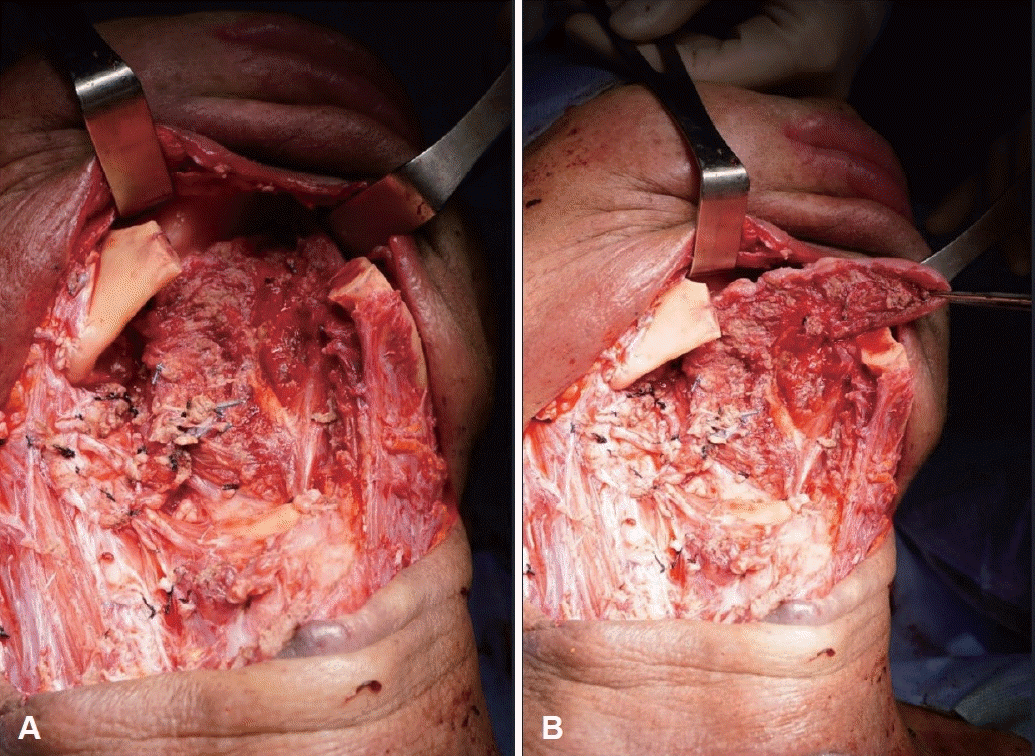
The salvage surgery was performed as follows: a gastrostomy performed by the gastrointestinal surgery team. After revision of neck dissection, using the visor flap approach, extensive resection of the oral cavity and oropharynx (tongue, floor of mouth, buccal space, retromolar trigone, and base of tongue), resection of sublingual space, right hyoid bone, and extrinsic tongue muscle, tooth extraction, wide segmental mandibulectomy (subtotal hemi-mandibulectomy), and submental skin resection were conducted. The resected mandible measured approximately 7 cm, with oral and oropharyngeal defects measuring approximately 15×7 cm and a submental skin defect of approximately 5 cm (Fig. 1). There appeared to be no suitable recipient vessel remaining for a free flap on the neck. Therefore, it was decided to use a pedicled flap.

Intraoperative appearance after extensive resection for recurrent oral and oropharyngeal cancer demonstrates mandibular defect of approximately 7 cm, oral and oropharyngeal defect of approximately 15×7 cm, and submental skin defect of approximately 5 cm. A: At rest. B: With protrusion of remained tongue.
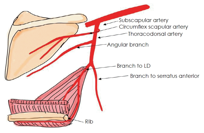
To cover the soft tissue defect, it was decided to use a LD flap with adequate volume. As for the accompanying bone flap, a choice had to be made between the scapular and rib. The scapular tip offers advantages in bone in-setting due to its three-dimensional structure, but its short rotating arc might make it difficult to reach the mandible, and there is also a risk of causing shoulder movement impairment. Therefore, it was decided to use the rib, which has a sufficient rotating arc and lower potential for functional impairment of donor site (Fig. 2).

Schema of the rib and latissimus dorsi (LD) muscle supplied by the descending branch of the thoracodorsal artery.
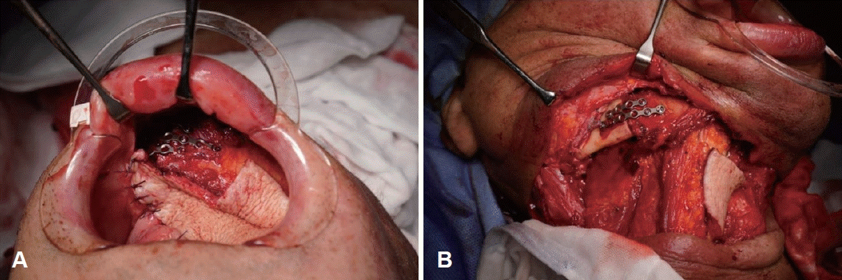
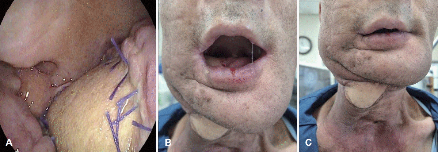
The surgical procedure proceeded as follows: A 20×7 cm right LD flap was planned for reconstruction of the oral cavity, oropharynx, and submental skin defects. Additionally, a 7 cm segment of the 9th rib, vascularized from the same thoracodorsal artery branch, was harvested together while maintaining its connection with the LD flap. The rib was dissected carefully, including some of the intercostal muscle, to ensure the safety of the supplying vessels and to prevent pneumothorax during dissection from the chest wall. After successfully obtaining the LD flap including the rib, a wide skin incision above the right clavicle was made to perform subcutaneous tunneling. This allowed the flap to be transferred from the donor site to the defect area. The mandible was reconstructed using the rib, while the LD flap was divided into two parts for reconstruction of the oral cavity, oropharynx, and submental skin (Fig. 3). There were no complications such as pneumothorax or flap and rib necrosis post-surgery, and the patient recovered well (Figs. 4 and 5).

Intraoperative view demonstrates using the rib to reconstruct the mandible, and dividing the latissimus dorsi flap into two parts to reconstruct the oral and oropharyngeal defect and the submental skin defect. A: Transoral view from above. B: External view from the right neck.

Postoperative appearance (A to C) at 1 month demonstrates well-healed flap without any necrosis. A: Intraoral view of the right oral cavity and oropharynx. B: External view of the face and neck with mouth open. C: External view of the face and neck with mouth closed.

Postoperative day 10 neck imaging shows proper alignment of the mandible with the rib. A: Neck enhanced CT image. B: T1 gadolinium enhanced image.
After 1 month, during a follow-up visit, it was confirmed that the surgical site had healed well with maintained mandible alignment properly. Subsequently, chemotherapy with platinum and pembrolizumab was initiated. However, evidence of recurrence was observed in the oral cavity 2 months postsurgery. Conventional chemotherapy was administered in response, but the recurrent cancer progressed, leading to the patient’s death 9 months after surgery.
Discussion
A review article published in 2023 aimed to establish the gold standard for mandibular reconstruction by analyzing 93 studies that met their criteria out of 15714 initially screened [1]. The most commonly used method identified was the fibular free flap, followed by iliac crest, scapula, and others. The study specifically analyzed cases where patients were unsuitable for free flap reconstruction, such as due to previous failed attempts, lack of suitable vessels in the neck due to prior chemotherapy and radiation therapy, or poor general medical condition. In such cases, the review analyzed outcomes of using titanium plates alone or in combination with regional flaps.
Using titanium plates offers advantages such as shorter surgical times, lower rates of donor site morbidity, and rapid functional rehabilitation compared to fibular free flaps [5]. However, it carries significantly higher risks of plate exposure and infection-related plate failures compared to fibular free flaps [7]. Given these drawbacks, the authors explored methods for reconstructing bone tissue using pedicled flaps alone, without resorting to titanium plates, particularly in patients unsuitable for free flap reconstruction.
The flap that was initially introduced for mandibular reconstruction was the free rib flap [9]. Following this, the LD-rib free flap has been reported in several studies, but unfortunately, it is now rarely used [1]. However, the authors have noted its potential to be used in the form of a pedicled flap and have confirmed that pedicled rib flap has been successfully used in recent spinoplastic reconstructions [10,11], and there have been successful case of mandibular reconstruction with pedicled LD-rib flap in the past [12].
In conclusion drawn from this case, the pedicled LD-rib flap can be effectively utilized when the use of fibular free flap is not feasible. This is due to its vascularized nature, which results in relatively shorter surgical times and higher survival rates compared to free flaps. Moreover, as it utilizes autologous bone similar to free bone flaps, it demonstrates better osteointegration and reconstruction success rates compared to titanium plates, which have higher exposure and infection rates [13,14]. To safely harvest the rib while maintaining its vascularity, some width of tissue including the intercostal muscle around the bone needs be preserved, attached to the bone, especially at the inferior, because the vessels travel along the bottom of the rib. The rib then should be cut off from the chest wall, maintaining the continuity of the pedicle. To cut the rib using saw, drill, or any other instrument, special care should be taken not to penetrate the pleura underlying, for example, by putting any kind of guard under the bone.
The scapula tip can also be used as a pedicled flap; however, it has limitations in terms of mobility compared to the rib and may lead to shoulder functional impairment [15]. The advantages of the LD-rib flap over chimeric flaps including scapular and LD are that the flap unit can be harvested at the same plane without additional incisions, it has the longer rotational arc, and the unit also can cover even similar large enough deficits. Another advantage of the pedicled LD-rib flap is its ability to obtain sufficient length of rib bone, enabling reconstruction of extensive mandibular defects [[14].
However, the pedicled LD-rib flap has several limitations compared to other free bone flaps. Firstly, unlike other free flaps which are advantageous for dental implantation, using pedicled LD-rib flap poses challenges in tooth implantation. Additionally, due to the substantial volume of soft tissue obtained from the LD muscle, reconstructive surgery can be difficult in cases where oral and cervical skin defects are not severe enough to require such volume. Moreover, ribs are thinner compared to other fibular flaps, making it challenging to contour them adequately to preserve the original angle of the mandible, which is important for aesthetic reasons.
Despite these limitations, the pedicled LD-rib flap still holds distinct advantages over other free bone flaps in mandibular reconstruction, making it sufficiently valuable. Therefore, it is considered a worthwhile approach to explore in future cases, given its strengths in mandibular reconstruction.
Notes
Acknowledgments
None
Author contributions
Conceptualization: Hye-Bin Jang, Joon Kyoo Lee. Data curation: all authors. Formal analysis: Hye-Bin Jang, Joon Kyoo Lee. Funding acquisition: Investigation: Hye-Bin Jang, Joon Kyoo Lee. Methodology: Hye-Bin Jang, Joon Kyoo Lee. Project administration: Hye-Bin Jang, Joon Kyoo Lee. Resources: all authors. Software: Hye-Bin Jang, Joon Kyoo Lee. Supervision: Joon Kyoo Lee. Validation: Joon Kyoo Lee. Visualization: Joon Kyoo Lee. Writing—original draft: Hye-Bin Jang. Writing—review & editing: Joon Kyoo Lee
