비배부 실리콘 임플란트로 인한 전두동 전벽 관통: 증례 보고
Penetration of Anterior Table of Frontal Sinus Due to Silicone Implant in Nasal Dorsum: A Case Report
Article information

Trans Abstract
A 33-year-old female with six prior augmentation rhinoplasties presented with a short, retracted nose and recurrent swelling over the nasal dorsum, without sinusitis symptoms. CT imaging revealed penetration of the anterior table of the frontal sinus by a silicone implant. Surgical intervention included implant removal, debridement of infected tissue, and reconstruction using the conchal cartilage and homologous fascia, tip grafting, and dorsum augmentation. Due to suspected infection, the planned costal cartilage reconstruction was postponed. In case of correcting the short nose resulting from silicone implantation, especially when infection is suspected, the risk of postoperative infection increases, potentially necessitating a second-stage operation. This case highlights the importance of comprehensive diagnostic methods, including CT imaging, and thorough preoperative consultation to ensure that patients understand potential surgical strategies and outcomes in revision rhinoplasty with complications from silicone implantation.
Introduction
Augmentation rhinoplasty often involves the use of synthetic materials such as silicone or Gore-Tex to enhance nasal structure and aesthetics. While these implants are generally considered safe, they may cause complications, especially when placed near the frontal sinus. Persistent pressure from the implant and the body’s inflammatory response can result in localized bone resorption or penetration [1]. Bone penetration of the anterior table of the frontal sinus is rare but significant, often leading to sinusitis, implant exposure, or infection. It is typically caused by mechanical factors, like pressure necrosis, or biological factors, such as chronic inflammation [2].
Correcting short noses in patients with prior silicone implants presents a challenge, especially with complications with a risk of infection. This case report describes an instance of anterior table penetration of the frontal sinus caused by a nasal implant, focusing on clinical presentation, diagnostic evaluation, and management strategy.
Case
A 33-year-old female with no significant comorbidities presented for revision rhinoplasty. Her primary concerns included dissatisfaction with her short, retracted nose and frequent swelling of the bony nasal dorsum. She denied symptoms of nasal obstruction, facial pain, rhinorrhea, hyposmia, anosmia, or frontal headache.
Her medical history revealed six prior augmentation rhinoplasties using homologous costal cartilage, silicone implant, the most recent performed 6 years earlier. She reported a substantial smoking history of one pack per day for 15 years but had ceased smoking and alcohol consumption 6 months before presentation.
On examination, her nose appeared short with a retracted columella, pinched nostrils, and an over-projected, upward-rotated tip, but no saddle nose deformity. The dorsum was smooth, with no swelling or definite fluctuation observed during the physical examination and the nostrils were asymmetrical (Fig. 1A). Nasal cavity examination showed no inflammatory signs.
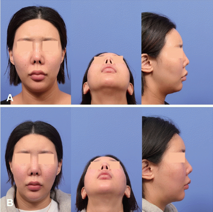
Preoperative facial images (A) and postoperative facial images at 5 months (B).
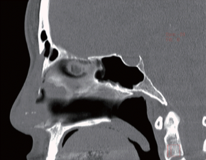
A CT scan revealed foreign body penetration of the anterior table of the frontal sinus without evidence of sinusitis (Fig. 2).
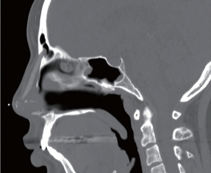
Sagittal CT scan cut showed penetration of anterior table of frontal sinus by a foreign body implant for a rhinoplasty.
The surgical plan included foreign body removal, revision rhinoplasty including reconstruction of septal framework, tip grafting, dorsum augmentation using costal cartilage, and reconstruction of anterior table defects of the frontal sinus. The patient was informed that if signs of infection were found during surgery, a second-stage operation would be needed, with the first surgery focusing on silicone removal and frontal sinus wall repair.
Under general anesthesia, a mixture of 2% lidocaine and 1:100000 epinephrine was infiltrated. Upon injection, a whitish, pus-like discharge was observed from the injection site of nasal dorsum. An inverted V-shaped trans-columellar incision with marginal incisions was performed. While exposing the nasal dorsum, a yellowish-green discharge was observed, and cultures were taken. The silicone implant was removed, and all purulent material was thoroughly suctioned.
The implant capsule was carefully dissected and debrided. While exposing of the upper lateral cartilages and nasal bones, a small defect on the left nasal bone was found, communicating with the nasal cavity (Fig. 3A). The periosteum was elevated to expose the frontal bone, which showed a defect approximately 1 cm in diameter (Fig. 3A). The defect on the anterior wall of the frontal sinus contained a small amount of granulation tissue but showed no evidence of pus (Fig. 3B). The granulation tissue was subsequently debrided. After its removal, the opening of the frontal sinus wall was identified (Fig. 3C).
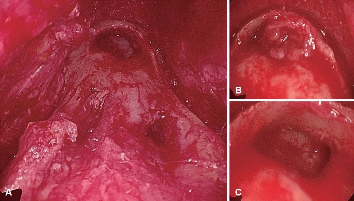
Intraoperative endoscopic findings of nasal bone and frontal sinus defects. A: Intraoperative endoscopic view of nasal dorsum showed left nasal bone defect and hole of anterior table of frontal sinus after removal of foreign body implant. B and C: Before debridement of granulation tissue in frontal sinus wall defect (B), after debridement (C).
The defects in the anterior table of the frontal sinus and left nasal bone were addressed by placing fascia and cartilage from the right auricular cymba concha to cover the frontal sinus defect, which was secured using Tisseel fibrin glue (Baxter Healthcare Corporation) (Fig. 4). The left nasal bone defect was reinforced with a homologous fascia, followed by the application of fibrin glue. Due to a highly suspected infection around silicone implant, the planned rhinoplasty for correction of short nose using costal cartilage was deferred to a later stage after complete closure of bony defect. Without separating the middle and medial crura of the lower lateral cartilage, the remaining harvested conchal cartilage was used for tip shield grafting. Irregularities in the bony dorsum, caused by nasal bone resorption due to the silicone implant, were smoothed using drilling. A 4 cm length of homologous fascia, consisting of five layers, was placed over the dorsum for augmentation.
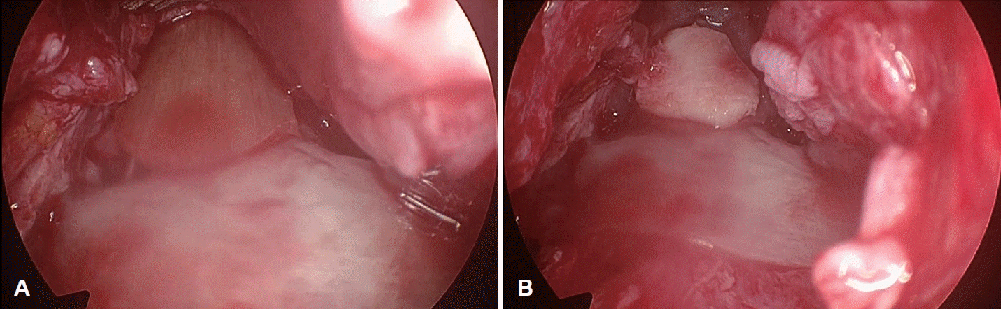
Reconstruction process of the nasal bone and frontal sinus defects. A: The defects was reconstructed by placing homologous fascia, secured with fibrin glue. B: Ear cartilage har- vested from the right auricular cymba concha, was placed on top of the fascia to reinforce the reconstructed site.
Postoperatively, microbiological cultures were negative for bacterial growth. A prophylactic dose of 1.5 g of intravenous ampicillin plus sulbactam was administered during anesthesia induction, followed by one week of oral antibiotics (amoxicillin plus clavulanic acid). There was no infection during the follow-up, and the patient was satisfied with the resolution of the recurrent swelling on dorsum and the correction of the nostril asymmetry, pinched nose deformity. However, the patient expressed a desire for further revision rhinoplasty to correct the insufficient tip projection, dorsal height (Fig. 1B).
Follow-up sagittal CT scans obtained approximately 2 months postoperatively confirmed that the reconstruction of the bony defect on the left bony dorsum, repaired with homologous fascia, ear cartilage, fibrin glue was stable. There were no signs of inflammation or fluid collection within the frontal sinus (Fig. 5).
Sagittal view of a CT scan obtained approximately 2 months postoperatively, demonstrating the reconstructed anterior table defect of the frontal sinus.
Discussion
This case highlights a rare complication of bone penetration in the anterior table of the frontal sinus due to foreign body implantation in rhinoplasty. Despite multiple prior surgeries and bone penetration, the patient showed no typical symptoms of sinusitis. However, CT revealed a defect in the frontal sinus wall, and surgery confirmed infection with significant inflammation. This underscores the importance of combining imaging methods, rather than relying solely on symptoms, for preoperative assessment. A thorough preoperative consultation is essential to offer appropriate surgical options tailored to the patient’s condition, as demonstrated in this case.
Short-nose deformities in revision rhinoplasty cases with a history of silicone implants present significant challenges. Corrective surgery typically involves nasal lengthening with septal reconstruction, dorsal augmentation, caudal rotation of the nasal tip with multilayer tip grafting, and elongation of the lateral compartment [3].
For lengthening the central compartment, the author prefers using a caudal septal extension graft placed between extended spreader grafts. In cases where dissection of the septal mucoperichondrium is difficult due to severe adhesions, a bypass L-strut graft, consisting of integrated costal cartilage for the columella and dorsal implant, may be used [3].
Revision rhinoplasty has a significantly higher risk of postoperative infection, about 20 times greater than primary surgery [4]. Our previous study found an infection rate of 3.39% with autologous costal cartilage grafting, mainly in revision cases for short-nose or saddle-nose deformities [4]. Contributing factors include large graft volumes disrupting nutrient diffusion, multiple fixation sutures causing irritation, and the rigid, scarred soft tissue envelope limiting accommodation of extensive grafting [4].
Furthermore, a smoking history has been identified as a predisposing factor for postoperative infection after rhinoplasty, likely due to the higher prevalence of Staphylococcus aureus colonization in smokers and the effects of various toxic substances that hinder wound healing, contribute to microvascular damage, and reduce oxygen delivery [5,6]. The patient’s smoking history was a contributing factor that increased the risk of infection after surgery.
Given intraoperative findings of pus discharge around the silicone implant and frontal sinus, nasal cavity communication, the surgical plan was modified to minimize infection risk. Instead of costal cartilage framework reconstruction, dorsum augmentation and tip grafting were performed using conchal cartilage and homologous fascia, avoiding medial and middle crura division.
Implant displacement is one of the most frequently observed issues. This is often attributed to the intrinsic properties of silicone, such as its soft texture and low friction [7]. However, implant migration can also result from iatrogenic factors during surgery. Contributing factors include improper prosthesis placement, dissection extending above the subperiosteal plane, asymmetrical nasal dorsum dissection, excessive osteotomy or dissection leading to bony dorsum damage, or uneven tissue contraction, possibly due to asymmetric soft tissue injury from previous rhinoplasty [8]. These factors can lead to gradual implant displacement within the dorsal pocket during healing.
In rare cases, intrusion into the anterior wall of the frontal sinus can occur due to the aforementioned iatrogenic factors, compounded by bone erosion associated with silicone implants. This erosion is driven by the formation of a tight capsule around the implant, the rigidity of the implant, and inadequate soft tissue coverage (thin flap) over the implant [9].
A case report documented a similar complication, emphasizing the importance of CT imaging and early intervention [2]. Unlike the typical findings of sinusitis or infection seen in cases of anterior table penetration, this patient exhibited no recurrent frontal sinusitis despite the presence of bone penetration. Beyond evaluating the patient’s symptoms, integrating preoperative CT scanning can provide a more comprehensive assessment of the patient’s condition, aiding in precise surgical planning.
In revision rhinoplasty for short-nose patients with suspected concurrent infection from silicone implant, it’s important to inform patients that intraoperative findings may differ from expectations, potentially requiring changes to the surgical plan. Patients should be counseled on the possibility of simultaneous or staged revision rhinoplasty to ensure a clear understanding of the surgical strategy and expected outcomes.
Notes
Acknowledgments
None
Author Contribution
Conceptualization: Yong Ju Jang. Data curation: Ammar Hussain Habibullah, Won Ki Cho. Resources: Yong Ju Jang. Supervision: Won Ki Cho. Writing—original draft: Ammar Hussain Habibullah. Writing—review & editing: Won Ki Cho, Yong Ju Jang.