A Case of Tumoral Calcinosis at Cervical Muscles
경부 근육에 발생한 종양성 석회증 1예
Article information

Abstract
Tumoral calcinosis is an uncommon ectopic calcification disorder that typically affects the periarticular soft tissue but rarely involves the cervical muscles. We report a 40-year-old male with an end-stage renal disease secondary to IgA nephropathy, who presented with a slowly enlarging right neck mass with tenderness and partial limitation of neck movement. Ultrasonography and contrast-enhanced CT demonstrated a multilobulated, densely calcified mass like lesion arising from the cervical muscles, displacing major neck vessels anteriorly. Serum studies revealed hyperphosphatemia with secondary hyperparathyroidism, which was diagnosed as secondary tumoral calcinosis. The mass was completely excised without complication. Histopathology showed amorphous calcified deposits surrounded by foreign-body giant cells. During 22 months of clinical and imaging follow-up, no recurrence has been observed. This case underscores the need to include tumoral calcinosis in the differential diagnosis of calcified cervical masses in dialysis patients in order that phosphate control and definitive surgery may achieve favorable outcomes.
Introduction
Soft-tissue calcifications in the head and neck generally reflect the nature of the involved structures and the mechanisms underlying calcium deposition. They may appear either as localized calcium deposits related to the anatomy and function of specific structures, such as the carotid artery or thyroid gland, or as localized manifestations in the head and neck of systemic pathologic processes. Although such softtissue calcifications are usually readily diagnosed on imaging, rare cases require additional evaluation, including serum studies and review of the medical history. One such example is tumoral calcinosis [1].
Tumoral calcinosis was first described by Inclan, et al. [2] in 1943. It presents as a calcified mass in the periarticular soft tissues, most commonly as multiple lesions involving the hip, elbow, shoulder, foot, and wrist [3,4]. Secondary tumoral calcinosis typically presents as a calcified periarticular mass associated with chronic renal failure. Because tumoral calcinosis rarely presents as a solitary lesion in the neck, diagnosis can be challenging [5-7]. Here, we report a rare case of secondary tumoral calcinosis arising in the right paravertebral neck region in a patient undergoing hemodialysis for end-stage renal disease caused by immunoglobulin A (IgA) nephropathy. To our knowledge, this presentation has not previously been reported in Korea. We also review the relevant literature.
Case
A 40-year-old male with a history of type 2 diabetes mellitus, hypothyroidism, gout, and end-stage renal disease secondary to IgA nephropathy had been receiving hemodialysis for 5 years. He presented with a progressively enlarging right neck mass that had been present for 2 months, accompanied by right-sided neck pain and limited neck motion. He had no history of trauma. On physical examination, a fixed, soft mass measuring approximately 5 cm was palpated in the right neck without overlying erythema or tenderness.
Ultrasound-guided fine-needle aspiration yielded a yellowish, caseous fluid. Neck ultrasonography showed a hyperechoic mass with an irregular margin and posterior acoustic shadowing (Fig. 1). Cytopathologic examination revealed calcified spherical structures and psammoma body-like calcifications. Preoperative contrast-enhanced CT revealed a 6-cm calcified, non-enhancing soft-tissue mass that appeared to arise from the right cervical paraspinal muscles. The mass extended from a point just lateral to the right transverse process of C1 to the right C4-5 facet region. At the C1 level, the mass also extended along the right side of the spinal canal and displaced the right internal jugular vein, internal carotid artery, and external carotid artery anteriorly (Fig. 2).
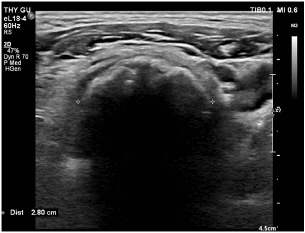
Neck ultrasonography. Irregular hyperechoic calcification with posterior acoustic shadowing.
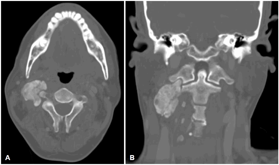
Neck CT scans show about 6 cm lobulated calcified mass in the right paravertebral space near the facet joint and transverse process, extending from the level of C1 to the level of C4/5disc space, suspicious to arise from the right paravertebral muscle, displacing Rt. IJV, ICA, and ECA anteriorly. A: Axial view. B: Coronal view. IJV, internal jugular vein; ICA, internal carotid artery; ECA, external carotid artery.
Serum chemistry showed an elevated creatinine level of 10.92 mg/dL (reference range [RR], 0.6 to 1.3 mg/dL), a phosphorus level of 7.3 mg/dL (RR, 2.5 to 4.9 mg/dL), a calcium-phosphate product (Ca*P) level of 75.92 mg2/dL2 (thresh hold, >55-60 mg2/dL2), and a parathyroid hormone level of 348.0 pg/mL (RR, 15 to 65 pg/mL). The serum calcium level was 10.4 mg/dL (RR, 8.6 to 10.6 mg/dL), total vitamin D was 35.53 ng/mL (RR, 30 to 50 ng/mL), and calcitriol was low at 5.66 pg/mL (RR, 18 to 72 pg/mL). Overall, these biochemical findings were consistent with secondary hyperparathyroidism and hyperphosphatemia.
Given the patient’s pain, limited neck motion, and the progressively enlarging lesion that displaced major vessels, surgical excision combined with conservative dietary modification was chosen. Intraoperatively, no gross neural or vascular invasion was observed. A multiloculated cystic mass containing yellowish, caseous material adherent to the surrounding muscles was identified and completely excised (Fig. 3). Macroscopically, the excised specimen was a multicystic mass measuring approximately 5.1×4.0×1.5 cm.
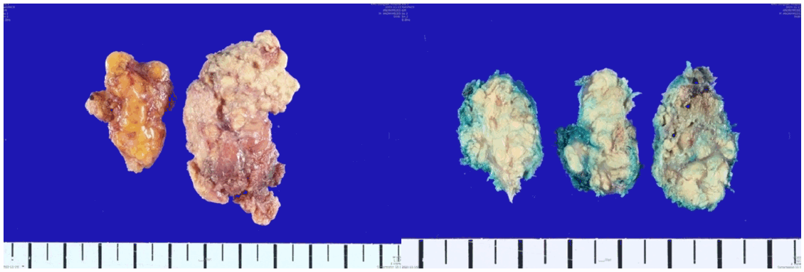
Gross findings of mass. It measures about 5.1×4.0×1.5 cm sized multiple cystic mass filled with cheese like material.
Postoperative histopathologic examination showed calcified nodular lesions surrounded by fibrous septa and inflammatory cells, including macrophages and osteoclast-like multinucleated giant cells. These findings confirmed the diagnosis of secondary tumoral calcinosis (Fig. 4). The patient showed improvement in neck pain and mobility after surgery and remained free of recurrence or complications during 22 months of follow-up.
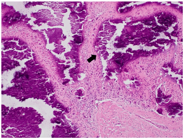
Pathologic slide view. Tumoral calcinosis with nodular lesions and fibrous septa. The nodule (arrow) shows an area of calcification surrounded by macrophages, osteoclast-lik e multinucleated giant cells, fibroblasts, and chronic inflammatory cells (H&E stain, original magnification ×100).
Discussion
Tumoral calcinosis is classified as either primary or secondary, and the primary form is further divided into normophosphatemic and hyperphosphatemic subtypes. Primary tumoral calcinosis typically presents in the second or third decade of life as a calcified soft-tissue mass. The normophosphatemic subtype is thought to be associated with SAMD9 mutations, whereas the hyperphosphatemic subtype is thought to involve mutations in GALNT3 and KLOTHO.
Secondary tumoral calcinosis usually presents as multifocal calcifications around large joints and is typically associated with chronic renal failure and dialysis. A decreased glomerular filtration rate leads to disordered phosphorus, calcium, and vitamin D metabolism, as well as secondary hyperparathyroidism. These metabolic abnormalities can increase the serum Ca*P, thereby increasing the risk of calcium deposition in extraosseous sites such as blood vessels and soft tissues [1,3].
Secondary tumoral calcinosis has rarely been reported in patients with chronic renal failure before the initiation of dialysis, and most cases occur in patients already undergoing dialysis. This may be because, when residual renal function is preserved, the serum Ca*P does not reach the 55-60 mg2/dL2 threshold considered necessary for the development of secondary tumoral calcinosis. Among patients undergoing hemodialysis or peritoneal dialysis, the reported prevalence of secondary tumoral calcinosis ranges from 0.5% to 3%. It usually develops after 2 years of dialysis, and the mean dialysis duration at diagnosis has been reported to be 6.47±3.17 years. Fewer than 10% of cases occur within 2 years after the initiation of dialysis, suggesting that cumulative phosphate burden during long-term dialysis may play an important role in the pathogenesis of this condition [8-10].
On imaging, CT typically shows periarticular amorphous, cystic, and multilobulated calcifications. A sedimentation sign may also be seen [4], in which high-density calcium deposits accumulate in the dependent portion of the lesion according to gravity [4]. Serum studies typically show elevated phosphorus, parathyroid hormone, and Ca*P levels, whereas calcium levels are normal or slightly elevated and 1,25-dihydroxyvitamin D levels are decreased. On histopathologic examination, amorphous calcium salt deposits are surrounded by inflammatory cells, including giant cells and mononuclear cells. These findings may help distinguish tumoral calcinosis from soft-tissue calcification secondary to bone destruction caused by other tumors or infection [7,9,11].
The primary treatment for secondary tumoral calcinosis is conservative medical management. Control of serum calcium and phosphorus levels is recommended through dietary phosphorus and calcium restriction and the use of low-calcium dialysate. Surgical treatment may be considered when medical treatment has failed or when mass effect or local invasion causes neurologic symptoms, limitation of joint motion, or a risk of injury to major structures. In patients with hyperparathyroidism that does not respond to medical treatment, subtotal or total parathyroidectomy may also be considered [1,7,9].
In the present case, the patient had chronic renal failure, a multilobulated calcified mass on CT, and elevated serum phosphorus, Ca*P, and parathyroid hormone levels. Histopathologic examination showed calcium salt deposition within multiple nodules, with peripheral aggregates of giant cells, macrophages, and chronic inflammatory cells. Taken together, these findings supported the diagnosis of secondary tumoral calcinosis. Given the patient’s pain, limited neck motion, and the relatively small, accessible location of the mass, we decided to perform surgical excision combined with conservative dietary modification.
In conclusion, secondary tumoral calcinosis typically presents as a calcified soft-tissue mass around large joints and may rarely arise in the neck. Most patients have a history of chronic renal failure and show characteristic imaging and laboratory findings. Depending on the clinical presentation, medical management is recommended as first-line treatment, whereas surgery may be considered when neurologic symptoms are present or when medical treatment has failed. Therefore, tumoral calcinosis should be included in the differential diagnosis of soft-tissue neck masses to facilitate appropriate initial treatment planning.
Supplementary Materials
Korean translation of this article is available with the Online-only Data Supplement at https://doi.org/10.3342/kjorl-hns.2026.00010.
Notes
Acknowledgments
None
Author Contribution
Conceptualization: Jae Man Lee, Soo Bin Bae, Chang Ki Yeo. Data curation: Jae Man Lee, Seong Chan Park, Soo Bin Bae. Formal analysis: Jae Man Lee. Investigation: Jae Man Lee, Seong Chan Park. Methodology: Jae Man Lee. Project administration: Jae Man Lee, Chang Ki Yeo. Resources: Jae Man Lee, Chang Ki Yeo. Software: Jae Man Lee. Supervision: Jae Man Lee, Chang Ki Yeo. Validation: Jae Man Lee. Visualization: Jae Man Lee. Writing—original draft: Jae Man Lee, Chang Ki Yeo. Writing—review & editing: all authors.