Simultaneous Perforation of the Lateral and Posterior Pharyngeal Walls Following Anterior Cervical Discectomy and Fusion: A Case Report
전방 경추 추간판 절제술 및 유합술 후 발생한 인두 측벽과 후벽의 동시 천공: 증례 보고
Article information

Abstract
Pharyngeal perforation is a rare but potentially fatal complication following anterior cervical discectomy and fusion (ACDF). Early recognition and prompt management are essential to prevent severe infections such as mediastinitis or sepsis. A 55-year-old man presented with sore throat and dysphagia one day after undergoing ACDF at another hospital. Laryngoscopy and neck computed tomography revealed a drainage tube penetrating the pharyngeal wall, accompanied by extensive subcutaneous emphysema and retropharyngeal abscess formation. Surgical exploration confirmed simultaneous perforations of the lateral and posterior pharyngeal walls, and primary repair of both defects was performed. The patient recovered without postoperative complications following surgical repair and antibiotic therapy. This case highlights the importance of early endoscopic and radiologic evaluation in patients presenting with dysphagia or subcutaneous emphysema after ACDF to ensure timely diagnosis and prevent life-threatening complications.
Introduction
Anterior cervical discectomy and fusion (ACDF) is a widely performed surgical procedure for the treatment of degenerative disc disease and myelopathy [1]. Although this procedure enables neural decompression and correction of cervical alignment, various complications can arise during the anterior cervical approach. Relatively common complications include dysphagia, hematoma, recurrent laryngeal nerve injury, and vocal cord paralysis, whereas pharyngeal or esophageal perforation is a rare but life-threatening complication [2-4].
If diagnosis and treatment are delayed, such injuries may progress to mediastinitis, sepsis, or death. They may also require extensive reconstructive surgery, thereby increasing morbidity and mortality [5]. However, because a sore throat and dysphagia immediately after surgery are common postoperative symptoms, they are easily overlooked, which makes early diagnosis of a pharyngeal injury difficult.
Pharyngeal perforation after ACDF is rarely reported in the medical literature. Here, we report a case of pharyngeal injury after ACDF, together with a review of the literature, to emphasize the importance of early diagnosis and treatment of pharyngeal injury.
Case
A 55-year-old man presented to our emergency department with a chief complaint of a sore throat that had developed one day earlier. He had a medical history of hypertension and had undergone C3-4 ACDF for cervical disc herniation at another hospital two days before presentation. Immediately after surgery, he developed a severe sore throat accompanied by dysphagia, odynophagia, hoarseness, a foreign body sensation, and exertional dyspnea. At presentation, his vital signs were normal except for a blood pressure of 160/100 mm Hg. Laboratory tests showed a white blood cell count of 13800/μL (neutrophils 85.8%) and a C-reactive protein level of 15.38 mg/dL.
Esophagogastroduodenoscopy performed at the outside hospital revealed a drainage tube and an anterior cervical screw fixation device protruding into the pharyngeal lumen (Fig. 1). Neck CT showed a drainage tube within the oropharynx (Fig. 2A), extensive emphysema in the left carotid space, submandibular space, parapharyngeal space, perithyroid space, and mediastinum (Fig. 2B), and fluid collection in the retropharyngeal space (Fig. 2C). Based on the clinical history and imaging findings, a pharyngeal wall laceration accompanied by a deep neck infection was suspected
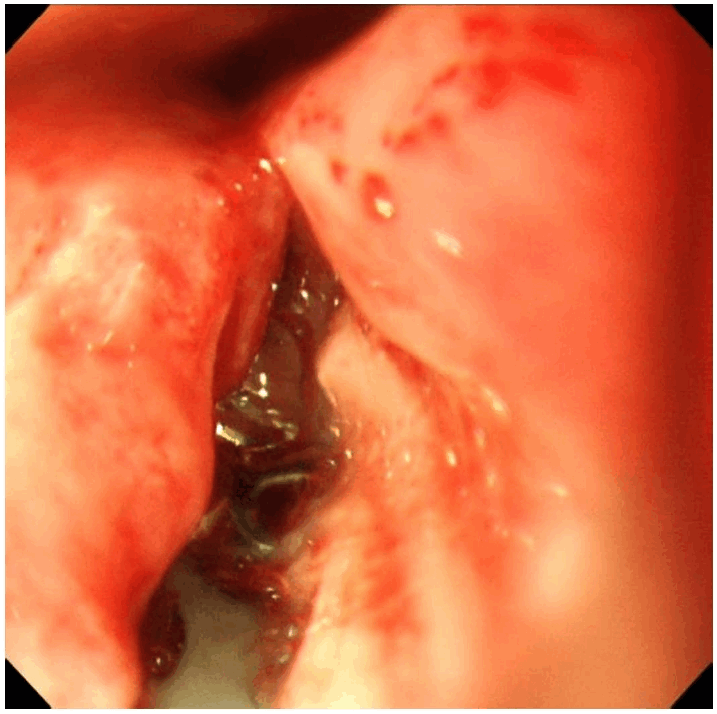
Upper esophagogastroduodenoscopy findings of the retropharyngeal injury after anterior cervical discectomy and fusion. The anterior cervical plate screw fixation hardware is visible within the oropharyngeal lumen.
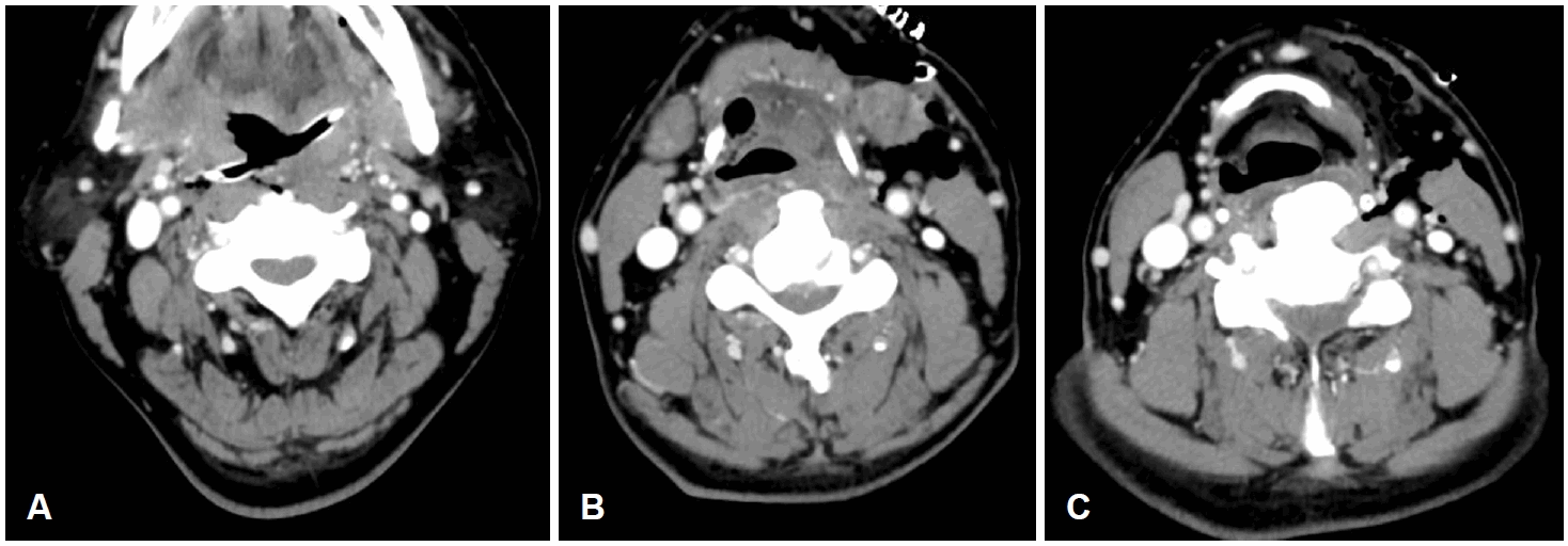
Preoperative contrast-enhanced neck CT images. A: A drainage tube is identified within the oropharyngeal lumen. B: Emphysema involving the parapharyngeal space, left carotid space and submandibular space. C: A fluid collection in the retropharyngeal space.
After endotracheal intubation under general anesthesia, the injured site was examined through a transoral approach. The drainage tube had penetrated the left lateral pharyngeal wall, entered the pharyngeal lumen, and extended toward the right side; however, no injury to the right pharyngeal wall was observed. An injury to the posterior pharyngeal wall extending from the oropharynx to the hypopharynx was confirmed. A cervical approach was required because adequate visualization of the hypopharyngeal region was difficult. The mechanism of injury was presumed to be penetration from the left lateral pharyngeal wall into the pharyngeal lumen, followed by perforation of the posterior pharyngeal wall during the insertion of the spinal instrument (Fig. 3)
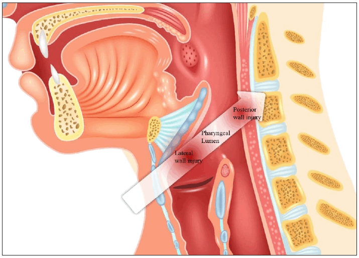
Schematic illustration of the mechanism of pharyngeal injury. During anterior cervical discectomy and fusion, the surgical instrument penetrated the lateral pharyngeal wall, traversed the pharyngeal lumen, and subsequently perforated the posterior pharyngeal wall before reaching the vertebral body during anterior cervical fixation.
Because the endotracheal tube prevented a clear evaluation of the posterior wall injury, a tracheostomy was performed, and the endotracheal tube was removed. Dissection was carried out along the previous drainage tube tract to the injured area on the lateral pharyngeal wall, which allowed direct visualization of the pharyngeal lumen and the prevertebral area adjacent to the posterior pharyngeal wall (Fig. 4A). Upon inspection, the posterior pharyngeal wall showed a vertical in cision extending from the mucosa to the muscular layer, without evidence of layer-by-layer dissection. Exposure of the anterior cervical screw fixation device was also confirmed. The operative field was evaluated intraoperatively in consultation with the orthopedic surgery team. No definite signs of infection were observed at the spinal operative site or around the anterior fixation screws. Accordingly, the fixation device was left in place without removal.
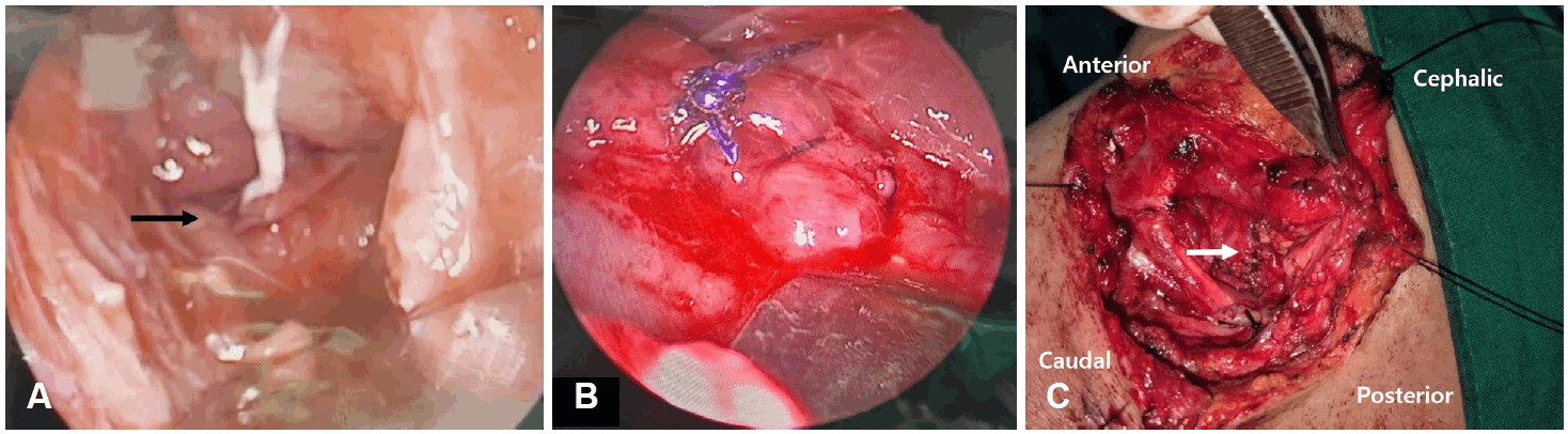
Intraoperative findings of the pharyngeal wall. A: Visualization of the prevertebral area adjacent to the posterior pharyngeal wall using a nasal speculum (black arrow). B: Primary closure of the posterior pharyngeal wall defect with combined muscular and mucosal layer sutures. C: Primary closure of the lateral pharyngeal wall defect with combined muscular and mucosal layer sutures (white arrow).
The injured mucosa and soft tissue were relatively clean with well-defined margins, and no marked inflammatory reaction or granulation tissue formation was observed. Therefore, primary closure was considered feasible. The defect in the posterior pharyngeal wall was repaired with five deep continuous sutures including the muscular and mucosal layers using a 4-0 barbed absorbable suture (V-Loc, Medtronic) (Fig. 4B). Because dissection of the prevertebral space was not extensive, no drain was inserted into the posterior pharyngeal injury site. The defect in the lateral pharyngeal wall was then closed with 4-0 Vicryl in the mucosal and submucosal layers so that the mucosal layer was appropriately inverted toward the lumen, followed by reinforcement with layer-by-layer suturing of the constrictor muscle (Fig. 4C). A drain was then inserted adjacent to the lateral pharyngeal wall to maintain drainage of the operative site.
Postoperatively, intravenous antibiotic therapy with teicoplanin and cefepime was started. Immediately after surgery, left-sided tongue weakness was observed, and hypoglossal nerve injury was suspected. However, because no definite transection of the hypoglossal nerve was identified intraoperatively, conservative treatment and close observation were undertaken. Initially, a small amount of drainage was noted through the drain inserted at the operative site, and the drainage volume gradually decreased. The drain was removed on postoperative day 8. Follow-up neck CT showed decreased emphysema and retropharyngeal fluid collection, along with improvement of the pharyngeal wall edema. The patient’s general condition improved, and he was discharged on postoperative day 15. At 2 months after surgery, the left-sided tongue weakness had improved, and he remains under outpatient follow-up without evidence of complications.
This study was approved by the Institutional Review Board (IRB No. BPIRB 2026-02-026).
Discussion
Esophageal perforation after ACDF is a rare but life-threatening complication, with a reported incidence of approximately 0.005% to 1.49% [6]. In contrast, reports of pharyngeal perforation are scarce in the literature, with most previously reported cases of pharyngeal injury describing single-site involvement [2,3,6]. Although dysphagia and hoarseness are commonly observed after surgery, an inadequately treated perforation can lead to mediastinitis, sepsis, or death.
Anatomically, the hypopharynx extends from the superior epiglottic margin to the inferior border of the cricoid cartilage at the C4-6 level. The Killian triangle at the C5-6 level and the lateral thyrohyoid membrane at the C3-4 level are vulnerable to injury due to their thin muscular layers [1]. Known primary causes include direct injury from retractors or surgical instruments, graft dislodgement, and injury during endotracheal intubation. Delayed perforations may also result from persistent pressure from implanted hardware or ischemic injury secondary to soft-tissue edema [7,8].
Clinically, symptoms usually appear early, most often immediately after surgery. Reported symptoms include dysphagia, fever, neck pain, and subcutaneous emphysema [2]. Subcutaneous emphysema is the most characteristic early sign and may be accompanied by crepitus on neck palpation [2]. When these symptoms occur, laryngoscopic examination and neck CT should be performed immediately to evaluate for pharyngeal or esophageal perforation.
For diagnosis, esophagography using a water-soluble iodinated contrast agent (e.g., Gastrografin) and CT are recommended as the initial imaging modalities. These modalities can evaluate the location of the perforation and the presence of mediastinal involvement [2]. If a water-soluble contrast study shows no leakage despite a strong clinical suspicion of perforation, barium esophagography may be cautiously considered due to its higher sensitivity [9]. However, barium leakage into the soft tissue or mediastinum can provoke foreign body or inflammatory reactions, necessitating careful patient selection based on the clinical situation.
Treatment is determined according to the size of the perforation and the clinical course. In cases of a localized small perforation without signs of infection, conservative treatment including nil per os, parenteral nutrition, and broad-spectrum antibiotics may be attempted [6]. Conversely, when the perforation is large or accompanied by complications such as inflammation or abscess formation, early surgical repair is required. The repair site may be reinforced using a muscle flap, such as the sternocleidomastoid or longus colli, or a free flap [10].
Reports of multiple lesions in the pharyngoesophageal region are exceptionally rare, with only a few cases identified to date. One case of multiple esophageal perforations associated with a deep neck infection was reported, and the patient recovered after reconstruction using colon interposition [11]. In another report, a bilateral hypopharyngeal injury after trauma was successfully treated with primary closure performed immediately after the injury [12]. Meanwhile, in a patient who underwent ACDF after cervical trauma, an esophageal perforation was identified approximately 20 days later. Primary closure was performed, but leakage persisted. Approximately 4 months later, multiple injuries of the lateral and posterior esophageal walls were identified, and the patient ultimately recovered after reconstruction using a thigh free flap [13]. Taken together, these cases suggest that multiple injuries in the pharyngoesophageal region can be difficult to manage with simple closure alone due to limited blood supply. However, if the injury is diagnosed early and treated appropriately, satisfactory outcomes can be expected with primary closure alone [12]. Conversely, when inflammation is severe, such as in a deep neck infection, or when diagnosis and treatment are delayed, the tissue condition deteriorates. Under these circumstances, reconstruction using a well-vascularized flap is more likely to be required [11,13].
Unlike previous reports that primarily describe single-wall involvement [2,3,6], the present case is a rare example of simultaneous injury to the lateral and posterior pharyngeal walls (Fig. 3). Despite this complex injury, treatment was performed early, and the tissue at the injured site remained relatively clean. Thus, recovery without complications was achieved with primary closure alone. This suggests that early evaluation and active intervention are important for improving the prognosis even in complex pharyngeal injuries after ACDF.
Supplementary Materials
Korean translation of this article is available with the Online-only Data Supplement at https://doi.org/10.3342/kjorl-hns.2026.00143.
Notes
Acknowledgments
None
Author Contribution
Conceptualization: Sung Yool Park. Data curation: Yanggyun Lee. Formal analysis: Yanggyun Lee, Sung Yool Park. Investigation: Yanggyun Lee, Do Hun Kim. Methodology: Yanggyun Lee, Sung Yool Park. Project administration: Sung Yool Park. Resources: Do Hun Kim, Tae Ui Hong, Sung Yool Park. Supervision: Sung Yool Park. Validation: Do Hun Kim, Sung Yool Park. Visualization: Yanggyun Lee. Writing—original draft: Yanggyun Lee. Writing—review & editing: Do Hun Kim, Tae Ui Hong, Sung Yool Park.