Simultaneous Tracheal and Esophageal Magnet Foreign Bodies in a Child: A Case Report
소아에서 발생한 기도 및 식도 동시 거치 자석 이물 1예
Article information

Abstract
Ingestion of multiple magnets is a growing concern as it often leads to severe complications like intestinal perforation or, rarely, tracheoesophageal fistula (TEF) due to magnetic attraction across tissue walls. We report a rare case of simultaneous tracheal and esophageal magnet foreign bodies found in a 9-year-old boy. Two adhered magnets were found across the trachea and esophagus, and six others in the intestine at day one of ingestion. To prevent acute desaturation, the tracheal magnet was prioritized and removed using a Storz ventilating bronchoscope. The esophageal magnet was subsequently identified and successfully extracted using a metallic suction tube, utilizing the property of magnets. This case emphasizes that early intervention is critical to prevent severe sequelae like TEF, as delayed management often results in fistula formation. A thorough understanding of magnet characteristics and surgical priority is essential for successful diagnosis and management of such complex foreign bodies.
Introduction
Foreign body ingestion is a common condition in children, occurring primarily between the ages of 6 months and 6 years [1]. In 80%-90% of cases, ingested foreign bodies pass spontaneously through the gastrointestinal tract without complications [2]. However, some may obstruct the airway or become lodged in the esophagus, requiring intervention [2]. The incidence of complications related to magnet ingestion has increased with the growing popularity of magnetic toys [3]. While ingestion of a single magnet typically does not cause significant problems, ingestion of multiple magnets, or a magnet together with another metallic object, can cause the objects to adhere to each other through magnetic forces, potentially leading to tracheoesophageal fistula (TEF), intestinal obstruction, intestinal fistulas, or perforation [4-6]. A TEF, in particular, can result from pressure necrosis when magnets become lodged simultaneously in the anatomically adjacent esophagus and airway, although this is very rare [6]. Here, we report a rare case of the successful removal of magnets that were simultaneously lodged in the airway and esophagus.
Case
A 9-year-old boy presented to our emergency department after ingesting multiple magnets. He was a normally developing child with no history of psychiatric disorders, such as autism spectrum disorder or attention deficit hyperactivity disorder, and no developmental delays. The day before presentation, the patient had accidentally swallowed several magnets. He was first evaluated at another hospital, where a plain chest radiograph showed two magnets in the bronchus, and an abdominal radiograph showed six additional magnets. His past medical history was unremarkable. On arrival, his vital signs were stable. His blood pressure was 110/60 mm Hg, pulse rate was 89 beats/min, respiratory rate was 20 breaths/min, and body temperature was 36.9°C.
Chest radiographs at our facility (Fig. 1A and B) revealed two metallic foreign bodies attached to each other within the mediastinum. One was located in the airway and the other in the esophagus. An abdominal radiograph (Fig. 1C) confirmed the presence of six metallic foreign bodies in the right lower quadrant of the abdomen. Given the risk of TEF formation and the potential for emergent complications, the decision was made to proceed with surgical removal. The procedure was planned in consultation with the thoracic surgery department, and the patient was placed under general anesthesia.
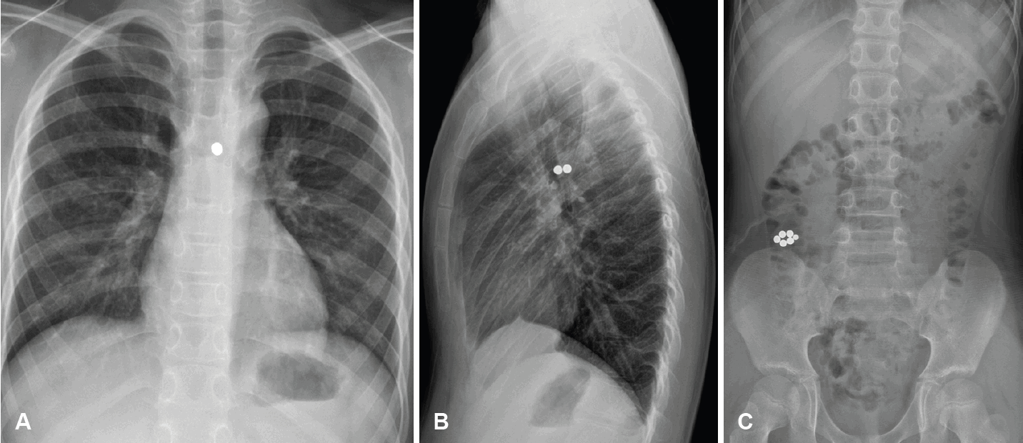
Preoperative simple radiographs. A: Posteroanterior chest radiograph showing a radiopaque cluster of spherical foreign bodies projected over the upper mediastinum. B: Lateral chest radiograph showing two round magnets adhering to each other across the posterior tracheal wall and anterior esophageal wall. C: Simple abdominal radiograph showing a cluster of six magnets in the right lower quadrant area.
Examination with a ventilating bronchoscope identified a round magnet in the left main bronchus (Fig. 2A). The magnet was removed using optical forceps (Fig. 3A). An ulcerative lesion was observed at the site of removal, but no definitive TEF was found (Fig. 2B). Immediate re-examination of the airway confirmed that no other foreign bodies remained. After the airway was secured with tracheal intubation, a bronchoscope of the same size was introduced into the esophagus, where a second round magnet was identified. A metallic suction tube (Fig. 3B) was advanced into the esophagus (Fig. 2C), and the magnet was successfully removed by allowing it to attach to the tube through magnetic attraction. The removed foreign body was confirmed to be a round magnet measuring 5 mm in diameter (Fig. 2D).
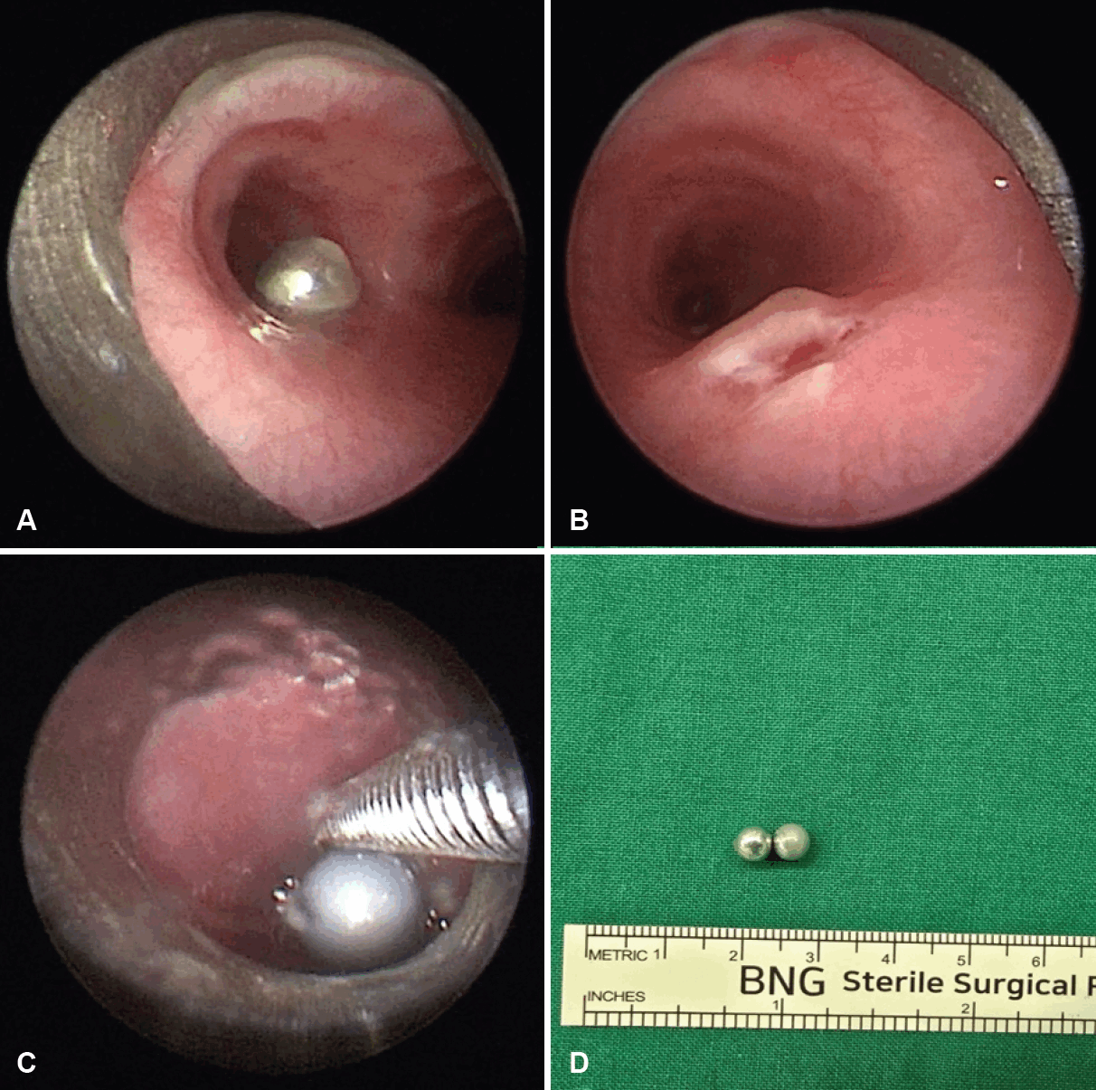
Intraoperative findings and the removed foreign bodies. A: Bronchoscopic view revealing a spherical magnetic foreign body in the left main bronchus. B: Mucosal ulceration and granulation tissue observed on the posterior bronchial wall after the removal of the foreign body. C: Esophagoscopic view showing the retrieval of the esophageal magnet using the magnetic attraction to a metallic suction tube. D: Two spherical magnets, each 5 mm in diameter, removed from the trachea and esophagus.

Surgical instruments used for the removal of foreign bodies. A: Optical forceps (KARL STORZ 10378 KF, Tuttlingen, Germany) used for the extraction of the tracheal magnets under bronchoscopic guidance. B: Rigid metallic suction tube (KARL STORZ 10381 B, Tuttlingen, Germany) utilized to retrieve the esophageal magnets by exploiting magnetic attraction.
Postoperatively, the patient was admitted to the Department of Pediatrics. A postoperative chest radiograph (Fig. 4A) confirmed removal of the two metallic foreign bodies from the esophagus and airway. A postoperative abdominal radiograph (Fig. 4B) showed that the six intra-abdominal magnets had migrated to the Lt. descending colon due to intestinal peristalsis. Prophylactic fasting was maintained for four days because the risk of delayed perforation or TEF formation from pressure necrosis of the mucosa could not be excluded. The passage of the six remaining intra-abdominal magnets was monitored with serial plain radiographs. Postoperatively, the patient received an intravenous proton pump inhibitor (PPI) to prevent the ulcer from worsening due to gastric acid reflux. Intravenous antibiotics were also administered for four days to prevent secondary infection. On the fourth postoperative day, upper gastrointestinal endoscopy was performed to assess healing of the esophageal mucosa and to rule out fistula formation. The examination revealed no abnormal findings, such as esophageal perforation, and oral feeding was initiated. The patient was asymptomatic at discharge, and no medications were prescribed.
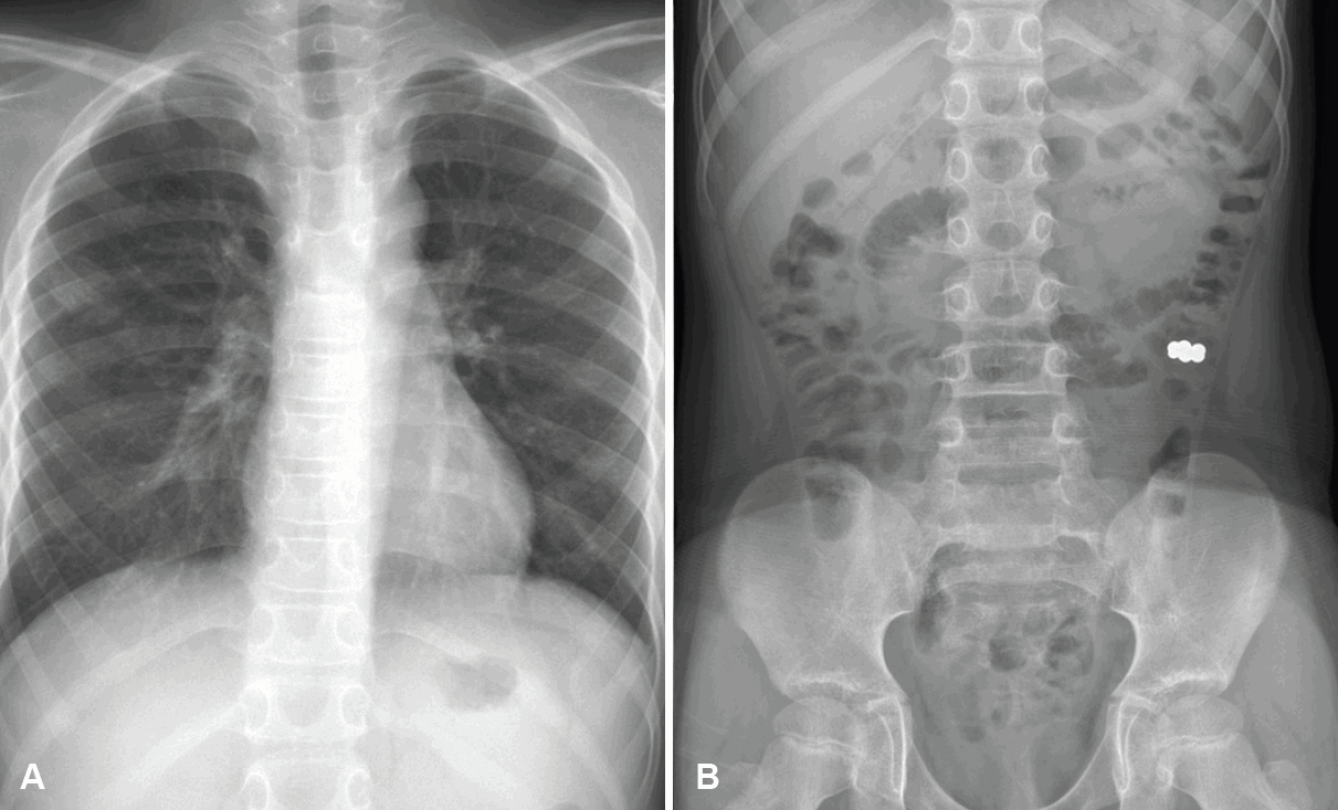
Postoperative simple radiographs. A: Posteroanterior chest radiograph showing the absence of foreign bodies in the trachea and esophagus. B: Simple abdominal radiograph showing the migration of the remaining magnets to the descending colon.
Discussion
Ingestion of foreign bodies is a very common occurrence in children [1]. Most of these objects pass spontaneously through the gastrointestinal tract without causing complications [2]. However, ingestion of multiple magnets can lead to serious complications due to magnetic attraction between them, including intestinal obstruction, intestinal fistulas, perforation, and TEF [4-6]. Diagnosis can be delayed because patients are often asymptomatic in the initial period following ingestion, which can sometimes lead to fatal outcomes [7].
Although this patient was older than the typical age for foreign body ingestion [1], this case is consistent with reports indicating that magnet ingestion occurs across a broad age range that includes adolescents [8]. This trend has been attributed to the distribution of magnets in various forms and an increase in cases where adolescents accidentally swallow or aspirate magnets while using them to simulate tongue or lip piercings [8]. Therefore, a high index of clinical suspicion for magnet ingestion is warranted for pediatric patients of all ages.
This case is highly unusual because the magnetic balls were both ingested and aspirated simultaneously. Similar cases of paired magnets lodged across the airway and esophagus have been reported by Ibrahim, et al. [9] and Ren, et al. [10]. In the case reported by Ibrahim, et al. [9], the primary symptom was acute respiratory distress resulting from airway compression caused by the attached magnets. Ren, et al. [10] reported a case in which a TEF had already formed as a result of pressure necrosis caused by the magnets.
In these complex situations, imaging is essential for treatment planning. Because attached magnets can appear as a single object, radiographs in at least two planes are necessary to accurately define their anatomical relationship [11]. In this case, the magnets appeared as a single object on the posteroanterior chest radiograph. However, the lateral view showed two magnets attached to each other, situated across the posterior airway wall and the anterior esophageal wall.
The foreign body in the airway was removed first to prevent it from being displaced distally by positive-pressure ventilation. Although the esophageal magnet could have migrated distally once the magnetic attraction was lost, the risk of it reattaching to the intra-abdominal magnets was considered low due to the significant anatomical distance. A 5 mm spherical magnet can be easily displaced distally during manipulation, so it was removed carefully using optical forceps. To remove the esophageal magnet, its magnetic properties exploited by allowing it to attach to a metallic suction tube. This technique was effective for minimizing both distal migration of the magnet and mucosal injury, and it may be a clinically useful strategy in similar cases.
The patient was kept fasting for 4 days postoperatively due to the confirmed pressure necrosis at the site. Similar to tissue injury from button battery ingestion, pressure injury from magnets carries a significant risk of delayed perforation or fistula formation, as tissue necrosis can continue even after the objects are removed [11]. Therefore, prophylactic fasting was implemented to minimize stress on the esophageal wall. Feeding was resumed only after upper gastrointestinal endoscopy on the fourth postoperative day confirmed the condition of the esophageal mucosa. An intravenous PPI was also administered to promote ulcer healing and prevent secondary injury from gastric acid reflux, in accordance with standard management guidelines for children with esophageal mucosal injuries [12].
The most critical difference between this case and those previously reported is the short time that elapsed between ingestion and treatment. In the case reported by Ren, et al. [10], more than one week had passed from ingestion to detection, and surgical correction was required for an already established TEF. In contrast, our patient presented within 24 hours of ingestion, which allowed for successful treatment via endoscopic removal alone. The distinct ulcerative lesion and inflammatory changes observed during the procedure (Fig. 2B) suggest that pressure necrosis in the mucosal layer had already begun within 24 hours of ingestion. Ultimately, early intervention prevented the formation of a severe fistula, reaffirming that prompt removal is the most critical prognostic factor in cases of magnet ingestion.
We have reported an uncommon case of the removal of magnets lodged simultaneously across the airway and esophagus in a child. A thorough understanding of the properties of magnetic foreign bodies is key to their successful diagnosis and treatment. Clinicians should be aware of the potentially fatal complications of multiple magnet ingestion, such as TEF and intestinal perforation. Prompt removal is critical for a favorable prognosis, and the appropriate use of metallic medical instruments based on the magnetic properties of the foreign bodies can be an effective removal strategy.
Supplementary Materials
Korean translation of this article is available with the Online-only Data Supplement at https://doi.org/10.3342/kjorl-hns.2026.00129.
Notes
Acknowledgments
This study was supported by a 2026 research grant from Pusan National University Yangsan Hospital.
Author Contribution
Conceptualization: Minhyung Lee. Data curation: Minhyung Lee, Jaemin Lee. Formal analysis: Jaemin Lee. Investigation: Jaemin Lee. Methodology: Jaemin Lee. Project administration: Jaemin Lee. Software: Jaemin Lee. Supervision: Eui-Suk Sung, Jin-Choon Lee, Minhyung Lee. Visualization: Jaemin Lee. Writing—original draft: Jaemin Lee. Writing—review & editing: all authors.