A Case of Seromucinous Hamartoma on the Natural Ostium of Maxillary Sinus
상악동 자연공에 발생한 장점액성 과오종 1예
Article information

Abstract
Seromucinous hamartoma (SMH) is rare among the hamartomas that occur from the sinonasal tract and the nasopharynx. Hamartomas can be categorized into seromucinous, respiratory epithelial adenomatoid, and chondromesenchymal subtypes. Among these, SMH have occasionally been observed in the posterior nasal septum and anterior nasal cavity. To date, however, there have been no documented cases originating from the natural ostium of the maxillary sinus. Herein, we describe a case of a 31-year-old man with SMH arising from the natural ostium of the left maxillary sinus. Although rare, this entity should be included in the differential diagnosis when evaluating mass lesions involving the maxillary sinus ostium.
Introduction
Hamartomas are benign, localized lesions composed of indigenous tissue elements that exhibit disorganized growth [1]. Occurrence within the nasal cavity is uncommon, and these lesions are broadly classified into three subtypes: seromucinous hamartoma (SMH), chondromesenchymal hamartoma, and respiratory epithelial adenomatoid hamartoma [2]. SMH consists of the proliferation of the respiratory epithelium and the subepithelial seromucinous gland [3]. Within the sinonasal tract, SMH most frequently involves the posterior nasal septum and nasopharynx, whereas involvement of the paranasal sinuses, lateral nasal wall, or anterior nasal cavity has been only rarely described [4-6]. In particular, origin from the natural ostium of the maxillary sinus is exceedingly uncommon, with no previously reported cases in the English-language literature. We report a distinctive case of a 31-year-old man with SMH arising from the natural ostium of the maxillary sinus.
Case
A 31-year-old man presented with symptoms of persisting rhinorrhea and foreign body sensation beginning after COVID-19 infections for more than 1 month. Nasal endoscopic examination at our outpatient clinic revealed a smooth, tanpink, polypoid mass occupying most of the left middle meatus (Fig. 1A). Further evaluation with computed tomography (CT) demonstrated a soft tissue lesion within the left nasal cavity extending to and obstructing the natural ostium of the maxillary sinus (Fig. 1B and C). The patient was subsequently admitted for surgical removal of the lesion.
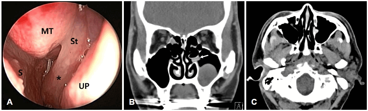
Endoscopic and radiologic findings of the nasal mass of the natural ostium of left maxillary sinus. A: Endoscopy showed a pinkish mass (star) with stalk presenting nodular surface in natural ostium of left maxillary sinus. B and C: Coronal and axial view of CT scan of the nose and paranasal sinuses showed the soft tissue density mass (white arrow) in natural ostium of left maxillary sinus. MT, denotes middle turbinate; UP, uncinate process; S, septum; St, stalk.
Under general anesthesia, endoscopic resection was performed using 0-degree and 30-degree nasal endoscopes. Intraoperatively, a tan-pink, polypoid mass was identified filling the natural ostium of the left maxillary sinus. The lesion was pedunculated, with its stalk attached to the margin of the natural ostium, and showed no additional attachments to surrounding structures within the nasal cavity or nasopharynx. The stalk was excised at its base, and the mass was removed using forceps (Fig. 2). Following excision, the natural ostium was widened. The entire specimen was retrieved and submitted for histopathological examination. Minimal bleeding was observed at the attachment site, with a feeding vessel noted within the stalk. Hemostasis was achieved with both absorbable and non-absorbable nasal packing, and the procedure was completed without complications.
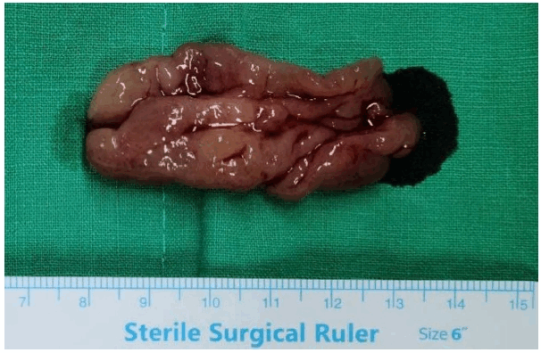
Macroscopic appearance of removed natural ostium seromucinous hamartoma presenting 5.5 cm tan-pink colored multinodular mass.
Histologically, the undulating surface of the mass was covered with ciliated, pseudostratified respiratory epithelium (Fig. 3A). Multifocal invagination of surface respiratory epithelium and lobules of seromucinous glands were seen in the stroma (Fig. 3B). The stroma was myxoid with inflammatory infiltration in superficial portion and fibrotic in deep portion. There was no nuclear atypia in epithelial cells.
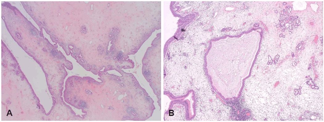
Histopathologic features of seromucinous hamartoma. A: The mass shows undulating surface with respiratory epithelial lining (H&E, ×10). B: Invagination of surface epithelium and seromucinous glands are seen in myxoid stroma (H&E, ×200). H&E, hematoxylin and eosin.
The postoperative course was uneventful, and the patient was discharged on postoperative day 2 without complications, including bleeding. He has since been followed regularly in the outpatient clinic, with no evidence of recurrence observed on nasal endoscopy during a 2-year follow-up period.
Discussion
Though the exact etiology of hamartoma is unknown, chronic inflammation to the nasal mucosa attributable to allergic rhinitis, bronchial asthma, or chronic rhinosinusitis is accepted as the most likely hypothesis [4]. SMHs have an equal sex distribution and a wide range of age distribution, though they are usually found in the sixth decade in the sinonasal tract [5]. In our case, unusually, the mass developed in his fourth decade.
When symptomatic, these lesions commonly present with nasal obstruction, rhinorrhea, or epistaxis. Sometimes it is found accidentally because they often have no symptoms [7]. In our case, he complained of unilateral rhinorrhea and foreign body sensation.
Macroscopically, SMH has a polypoid appearance and may have a broad base or a stalk [7]. Sinus Endoscopy showed a tanpink, polypoid mass filling the left middle nasal meatus. Its morphology closely resembled that of an antrochoanal polyp. Given that antrochoanal polyps generally arise from the osteomeatal complex, this entity was considered the most likely preoperative diagnosis. Intraoperatively, however, the lesion was identified as a pedunculated mass originating from the natural ostium of the maxillary sinus.
The differential diagnosis for SMH includes low-grade sinonasal adenocarcinoma, respiratory epithelial adenomatoid hamartoma, and inflammatory nasal polyps. The distinction between SMH and the inflammatory polyp is important because inflammatory polyps usually have recurrence rates from 10% to 30%, while SMH usually does not relapse [7].
From a histopathologic standpoint, inflammatory polyps typically lack epithelial proliferation, although glandular elements may occasionally be present. In addition, the stromal background of inflammatory polyps is characteristically edematous and loosely textured, with a mixed inflammatory infiltrate rich in eosinophils [7].
Radiologic evaluation, including CT or magnetic resonance imaging, can aid in assessing lesion size, extent, and local involvement. Immunohistochemical studies may further support the diagnosis; SMH is typically positive for S100, CK7, and CK19, and demonstrates a near absence of basal cells [8].
A current standard treatment of sinonasal SMH is endoscopic mass excision. To the best of our knowledge, there has been only one reported recurrence case after resection [9,10].
To the best of our knowledge, this represents the first reported case of SMH arising from the natural ostium of the maxillary sinus. Although extremely uncommon, SMH should be considered in the differential diagnosis of lesions in this location, and all excised tissue from the maxillary sinus ostium should undergo histopathological evaluation to ensure accurate diagnosis.
Notes
Acknowledgments
None
Author Contribution
Conceptualization: Seong Kook Park. Data curation: Seong Kook Park. Investigation: Seong Kook Park. Methodology: Seong Kook Park. Resources: Seong Kook Park. Supervision: Tae Ui Hong. Writing—original draft: Soohyun Sim, Eung Rok Yeon. Writing—review & editing: Tae Ui Hong, Seong Kook Park.