Intratympanic Corticosteroid Therapy for Treatment-Resistant Otitis Media With Effusion: A Systematic Review and Meta-Analysis
Article information

Trans Abstract
This systematic review and meta-analysis evaluated the efficacy and safety of intratympanic (IT) corticosteroid therapy compared with that of standard management of patients with treatment-resistant otitis media with effusion (OME). PubMed, Scopus, Web of Science, and the Cochrane Library were searched from inception to March 2026. Randomized controlled trials and observational studies comparing IT corticosteroid therapy with standard treatment were included. The primary outcomes were changes in pure-tone average (PTA) hearing thresholds and Type A tympanogram normalization. Secondary outcomes included air-bone gap improvement, OME recurrence, tympanosclerosis, otorrhea, and tympanic membrane perforation. Pooled effect estimates were calculated using random-effects models. Six studies involving 312 patients and 417 evaluated ears were included. Overall, IT corticosteroid therapy significantly improved the PTA hearing thresholds compared with the standard treatment (mean differences [MD]=-5.57 dB; 95% confidence interval [CI], -8.15 to -2.99; p<0.001; I²=71.88%). Stratified analyses showed significant improvements in both pediatric patients receiving IT methylprednisolone plus ventilation tube insertion (VTI) (MD=-4.03 dB; 95% CI, -4.98 to -3.07) and in adult patients receiving IT dexamethasone without VTI (MD=-13.68 dB; 95% CI, -19.52 to -7.84). IT corticosteroid therapy also significantly improved the air-bone gap (MD=-7.06 dB; 95% CI, -10.94 to -3.17), reduced the OME recurrence (risk ratios [RR]=0.27), and reduced tympanosclerosis (RR=0.21). No significant differences were observed in Type A tympanogram normalization, otorrhea, or tympanic membrane perforation. IT corticosteroid therapy may improve hearing outcomes and reduce recurrence in patients with persistent or treatment-resistant OME with no statistically significant increase in treatment-related adverse events detected. However, larger size trials with patient-level analyses, standardized protocols, and clinically meaningful hearing outcomes are required.
Introduction
Otitis media with effusion (OME) refers to the presence of fluid in the middle ear without the clinical signs or symptoms of acute middle ear infection [1,2]. OME represents one of the most common causes of hearing impairment in children, with more than 90% of children experiencing at least one episode before the age of 10 years, most commonly between the ages of 6 months and 4 years [3]. The pathogenesis of OME is multifactorial and primarily associated with eustachian tube dysfunction, leading to impaired middle ear ventilation, negative pressure, and subsequent fluid accumulation within the tympanic cavity [4]. Additionally, immunological and inflammatory processes contribute to the development and persistence of middle ear effusion [5]. Early and appropriate treatment of OME is important to prevent hearing impairment, speech delay, and developmental problems in children. Current guidelines from the American Academy of Otolaryngology-Head and Neck Surgery (AAO-HNS) recommend myringotomy with ventilation tube insertion (VTI) for children with persistent OME (>3 months) or for groups with high risk such as those with cleft lip/palate or other craniofacial anomalies [6]. However, simple myringotomy with aspiration of middle ear effusion alone has limited therapeutic value because the incision usually heals within a few days, while VTI may be associated with complications such as tube otorrhea [7-9].
Steroids are biologically active compounds with potent antiinflammatory properties and have been investigated for OME because they may reduce middle ear inflammation and mucin production and improve Eustachian tube function [10]. Although systemic and intranasal steroids have been proposed as alternatives to surgery, previous evidence has not demonstrated sustained benefit for effusion resolution or hearing outcomes [11]. Recently, intratympanic (IT) steroid administration has gained more attention as a more effective delivery method, providing high local drug concentrations in the middle ear while minimizing systemic absorption [12]. Previous studies have suggested beneficial effects of IT steroid therapy. A study comparing IT steroids to placebo in children over 12 and adults with OME demonstrated improvements in symptoms and quality of life for the steroid group [13]. Similarly, another study investigating the use of IT steroids as an adjunct to myringotomy and VTI in children reported lower rates of symptom recurrence and postoperative complications [14]. However, the evidence remains limited and clinically heterogeneous. Therefore, this systematic review and meta-analysis evaluated the effectiveness and safety of IT corticosteroid therapy compared with standard management in patients with persistent or treatment-resistant OME.
Method
This systematic review was conducted in accordance with the Preferred Reporting Items for Systematic Reviews and Meta-Analyses (PRISMA) guidelines [15]. The methodology adhered to the recommendations outlined in the Cochrane Handbook for Systematic Reviews of Interventions [16].
Data sources and search strategy
A comprehensive literature search was conducted across major electronic databases, including PubMed, Scopus, Web of Science, and the Cochrane Library from inception to March 2026, to identify relevant studies evaluating the use of IT steroids compared with standard treatment in patients with OME resistant to conventional medical treatment. The search strategy was developed using Boolean operators and incorporated the following search terms: (“otitis media with effusion” OR “OME”) AND (“intratympanic steroid” OR “intratympanic corticosteroid” OR “dexamethasone” OR “methylprednisolone” OR “budesonide”). Only studies published in English were considered in search. Additionally, forward and backward citation analysis was performed by screening the reference lists of the included articles and reviewing studies that cited them to identify any additional relevant publications. The detailed search strategy tailored for each database is provided in the Supplementary Table 1.
Study selection
All records identified through the database searches were imported into EndNote reference software, for removal of duplicates. The remaining articles were then uploaded to the Rayyan platform for further assessment [3]. Tworeviewers independently screened the titles and abstracts of the retrieved studies to identify potentially eligible articles. Full text of the selected studies were subsequently assessed in detail for eligibility based on the predefined PICO criteria. Reviewer’s discrepancies were addressed through discussion, and when needed, a third reviewer was consulted.
Eligibility criteria and outcomes
Studies included if they were randomized controlled trials (RCTs) or observational studies that evaluated the effectiveness of IT steroid therapy in addition to standard treatment in patients with OME resistant to conventional medical treatment, compared with standard treatment alone. The interventions in the included studies involved different types of IT corticosteroids, including methylprednisolone, dexamethasone, and budesonide. Standard treatment consisted of either surgical management with VTI alone following myringotomy without IT steroid administration, or conventional medical therapy, including saline nasal irrigation, oral antibiotics, intranasal corticosteroids, oral antihistamines, systemic steroids, and topical or systemic decongestants such as pseudoephedrine and xylometazoline nasal spray. The primary outcomes were change in pure-tone average (PTA) hearing threshold and Type A tympanogram normalization. Secondary outcomes included OME recurrence, tympanosclerosis, otorrhea, tympanic membrane perforation, and air-bone gap improvement.
Data extraction and risk of bias assessment
Data was extracted separately by two reviewers using a standardized form, capturing study characteristics (study ID, country, study design, time frame, and follow-up duration), sample size, data of intervention and control therapies, participant demographic characteristics (mean age and gender distribution), study inclusion criteria, main conclusions reported by the authors, and outcome measures. The risk of bias of the included RCTs was assessed using the Cochrane Risk of Bias tool version 2 (RoB-2) [17]. Observational studies were assessed using the Newcastle-Ottawa Scale (NOS) [18], which assess study quality depending on selection of participants, comparability of study groups, and assessment of outcomes. Reviewer’s discrepancies were addressed through discussion, and when needed, a third reviewer was consulted.
Statistical analysis and heterogeneity assessment
Statistical analyses were performed using STATA software version 19.5 SE (StataCorp). For dichotomous outcomes, pooled risk ratios (RRs) with 95% confidence intervals (CIs) were calculated, while continuous outcomes were pooled as mean differences (MDs) with 95% CIs. A random-effects DerSimonian-Laird model was used to account for anticipated clinical and methodological heterogeneity. The unit of analysis was determined according to the original study report. Patient numbers were used when outcomes were reported at the patient level, whereas ear numbers were extracted when outcomes were reported at the ear level. In split-ear study designs, where one ear received IT steroid therapy and the contralateral ear served as the control, outcomes from each ear were extracted according to the allocated treatment group. Statistical heterogeneity was assessed using Cochran’s Q test (p<0.10 indicating significant heterogeneity) and quantified using the I² statistic, where values of 25%, 50%, and 75% represented low, moderate, and high heterogeneity, respectively. Between-study variance was estimated using τ². The robustness of the pooled estimates was evaluated using sensitivity analyses with a leave-one-out approach.
Subgroup analyses were conducted for the primary outcomes to explore potential sources of clinical and methodological heterogeneity. For PTA, age group, corticosteroid type, and VTI strategy formed the same 2-versus-2 partition: the pediatric studies evaluated IT methylprednisolone plus VTI, whereas the adult studies evaluated IT dexamethasone without VTI. These variables were therefore collapsed into one exploratory comparison of bundled clinical profiles rather than analyzed as independent moderators. A separate exploratory analysis compared RCTs with nonrandomized studies. For Type A tympanogram normalization, age and VTI strategy were also collinear and were represented by a single exploratory clinical-profile comparison. Additional Type A analyses according to corticosteroid type and study design were regarded as hypothesis-generating.
Results
Literature search
A total of 404 records were identified through database searching. After the removal of 47 duplicate records, 357 studies remained for title and abstract screening. Of these, 338 records were excluded as irrelevant, leaving 19 reports for full-text assessment based on the predefined PICO criteria. Ultimately, six studies met the inclusion criteria and were included in the systematic review and meta-analysis [13,14,19-22]. The study selection process is shown in Fig. 1.
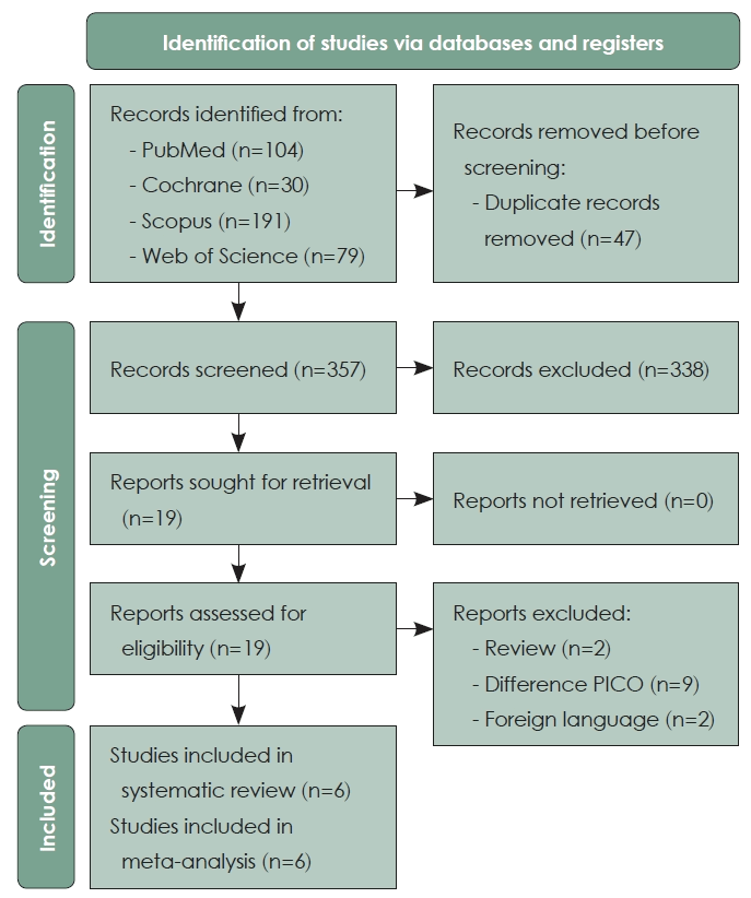
PRISMA flow diagram.
Characteristics of the included studies
A total of six studies were included in this review, comprising four RCTs and two non-randomized controlled studies. Overall, the included studies enrolled 312 patients. Because several studies reported outcomes at the ear level rather than the patient level, a total of 417 evaluated ears were available across the included studies. Of these, 239 ears received IT steroid therapy, while 178 ears received standard treatment. The follow-up duration ranged from 1 to 24 months, with a mean of 7 months and a median of 3 months. The included population consisted of pediatric and adult patients with OME resistant to conservative treatment. The intervention group received IT steroid injections (methylprednisolone, dexamethasone, or budesonide), whereas the control group received standard management. A detailed summary of the studies included is presented in Table 1.
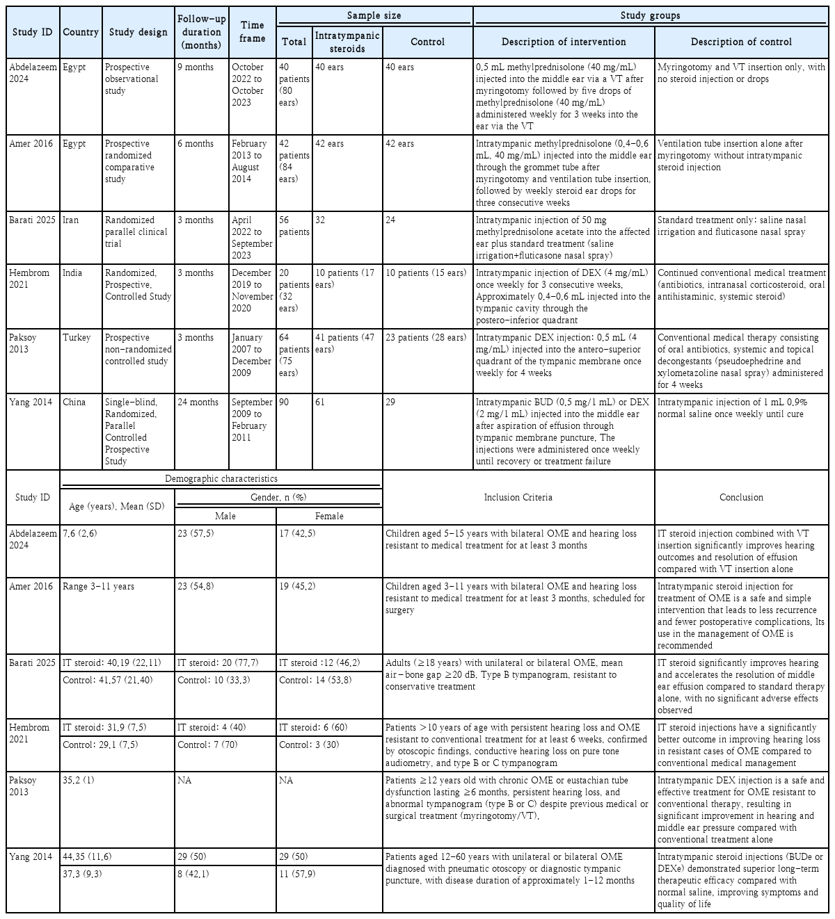
Summary and baseline characteristics of the included studies
Risk of bias assessment
The methodological quality of the included studies was assessed using appropriate tools according to the study design. RCTs were evaluated using the RoB-2 tool, with some studies judged as having a low risk of bias and others presenting some concerns. Cohort studies were evaluated using NOS and were considered to be of high methodological quality (Supplementary Figs. 1 and 2).
Primary outcomes
PTA hearing threshold improvement
The change in PTA hearing threshold from baseline was evaluated across four studies comparing IT steroid therapy with standard treatment. The overall random-effects DerSimonian-Laird model showed that IT steroid therapy was associated with a significant improvement in PTA compared with the control group (MD=-5.57 dB; 95% CI, -8.15 to -2.99; p<0.001; I²=71.88%). However, substantial heterogeneity was observed across the included studies (τ²=3.61; I²=71.88%). To explore this heterogeneity, stratified clinical-profile analyses showed statistically significant improvements in PTA in both clinical settings. Among pediatric patients, IT methylprednisolone administered as an adjunct to VTI was associated with a greater reduction in PTA threshold than VTI alone (MD=-4.03 dB; 95% CI, -4.98 to -3.07; I²=0%). Among adult patients, IT dexamethasone administered without VTI was associated with a greater reduction in PTA threshold than standard medical treatment (MD=-13.68 dB; 95% CI, -19.52 to -7.84; I²=0%) (Fig. 2). A leave-one-out sensitivity analysis was performed to evaluate the robustness of the pooled estimate. The overall treatment effect remained statistically significant after the sequential omission of each individual study, indicating that no single study disproportionately influenced the pooled result (Supplementary Fig. 3). In the exploratory study-design analysis, the pooled effects favored IT corticosteroid therapy but were not statistically significant among RCTs (MD=-9.14 dB; 95% CI, -20.70 to 2.43; I²=83.77%; p=0.12) or nonrandomized studies (MD=-7.20 dB; 95% CI, -15.13 to 0.74; I²=77.79%; p=0.08). The between-design difference was not significant (Qb[1]=0.07; p=0.79) (Supplementary Fig. 4).
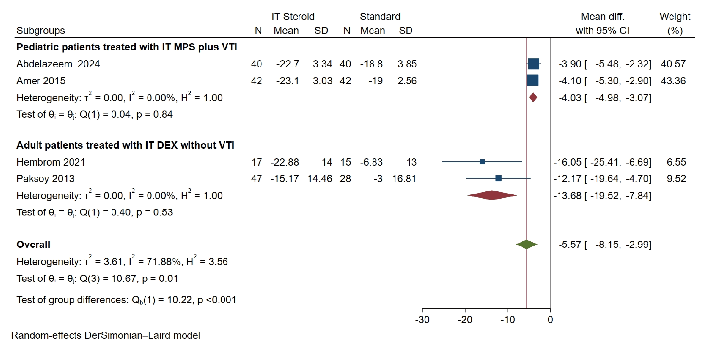
Stratified forest plot of PTA improvement in pediatric patients treated with IT MPS+VTI and adult patients treated with IT DEX without VTI. PTA, pure-tone average; IT, intratympanic; CI, confidence interval; VTI, ventilation tube insertion; MPS, methylprednisolone; DEX, dexamethasone.
Type A tympanogram normalization
The rate of Type A tympanogram normalization was evaluated across five studies comparing IT steroid therapy with standard treatment. The pooled effect estimate using a random-effects DerSimonian-Laird model showed that IT steroid therapy was not significantly associated with Type A tympanogram normalization compared with standard treatment (RR=1.25; 95% CI, 0.94 to 1.65, p=0.12). Low heterogeneity was observed among the included studies (τ²=0.02, I²=19.01%) (Fig. 3). A leave-one-out sensitivity analysis showed that the overall effect remained non-significant after sequential omission of each individual study (Supplementary Fig. 5). In the exploratory bundled-profile comparison, the RR was 1.10 (95% CI, 0.78 to 1.55) for pediatric patients receiving IT corticosteroid plus VTI and 1.40 (95% CI, 0.93 to 2.11; I²=29.85%) for adolescent/adult patients receiving IT corticosteroid without VTI; the between-profile difference was not significant (Qb[1]=0.81; p=0.37) (Fig. 4). A separate subgroup analysis according to steroid type showed no significant improvement in the budesonide-, dexamethasone-, or methylprednisolone-based studies (RR=1.36, 95% CI, 0.71 to 2.59; RR=2.10, 95% CI, 0.77 to 5.74; and RR=1.09, 95% CI, 0.82 to 1.44, respectively) (Supplementary Fig. 6). Subgroup analysis according to study design also showed no significant improvement in either RCTs or non-randomized studies (Supplementary Fig. 7).
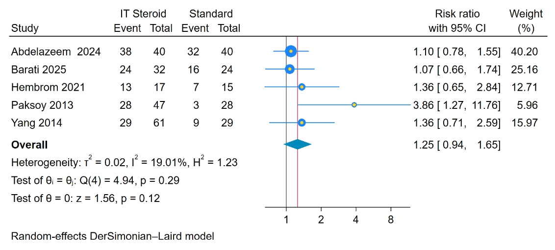
Forest plot of Type A tympanogram normalization. IT, intratympanic; CI, confidence interval.
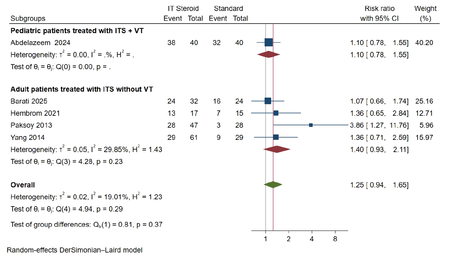
Exploratory subgroup analysis of Type A tympanogram normalization comparing pediatric patients treated with ITS+VT versus adolescent/adult patients treated with ITS without VT. IT, intratympanic; CI, confidence interval; ITS, intratympanic steroids; VT, ventilation tube.
Secondary outcomes
Clinical outcomes
Pooled analysis demonstrated that IT steroid therapy significantly decreased the recurrence of OME compared with standard treatment (RR=0.27, 95% CI, 0.09 to 0.78; p=0.01, I²=0%) and reduced the risk of tympanosclerosis (RR=0.21, 95% CI, 0.05 to 0.83; p=0.03, I²=0%). In contrast, pooled analysis showed no significant difference between IT steroid and standard treatment regarding otorrhea (RR=0.71, 95% CI, 0.31 to 1.66; p=0.43, I²=0%) or tympanic membrane perforation (RR=0.62, 95% CI, 0.14 to 2.80; p=0.54, I²=0%) (Supplementary Figs. 8-11).
Audiological outcome (air-bone gap)
Pooled analysis using a random-effects model showed that IT steroid therapy significantly improved the air-bone gap compared with standard treatment (MD=-7.06, 95% CI, -10.94 to -3.17; p<0.0001), with no heterogeneity observed (I²=0%) (Fig. 5).
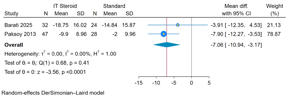
Forest plot showing the effect on air–bone gap. IT, intratympanic; CI, confidence interval.
Discussion
This meta-analysis offers an updated overview of the available evidence on IT corticosteroid therapy for persistent or treatment-resistant OME. The findings suggest that IT corticosteroid therapy may improve audiological outcomes, in-cluding PTA hearing thresholds within two distinct clinical profiles and the pooled ABG, while also reducing OME recurrence and tympanosclerosis. These potential benefits were not accompanied by significant increases in treatmentrelated adverse events, including otorrhea and permanent tympanic membrane perforation. However, Type A tympanogram normalization was not statistically significantly improved, indicating that functional hearing recovery may not necessarily correspond to complete physiological restoration of middle ear ventilation.
The biological mechanisms underlying the efficacy of IT steroids involve multi-targeted anti-inflammatory and physiological actions within the middle ear. OME is characterized by secretory transformation of the middle ear epithelium, subepithelial edema, and the infiltration of inflammatory cells [21]. IT steroids act by inhibiting the arachidonic acid cascade, thereby reducing the production of inflammatory mediators and lipopolysaccharides known to induce middle ear effusion [22]. Furthermore, steroids are noted to inhibit mucin production and enhance Eustachian tube function by promoting the secretion of surfactants and improving the transport capacity of the ciliary epithelium [13,23,24]. This direct application allows for higher local drug concentrations compared to systemic delivery, which often fails to achieve therapeutic levels in the middle ear space without significant systemic side effects [12,14].
Eosinophilic otitis media should be considered in patients with recurrent or treatment-resistant middle ear effusion. This condition is characterized by eosinophil-rich, often highly viscous middle ear effusion and is commonly associated with bronchial asthma and chronic rhinosinusitis with nasal polyps [25,26]. It has been shown to respond well to corticosteroid therapy; therefore, IT corticosteroid administration may reduce local eosinophilic inflammation and improve middle ear symptoms [27].
To explore the heterogeneity observed in the PTA analysis, we combined the overlapping subgroup analyses into a single exploratory comparison between pediatric patients treated with IT methylprednisolone (MPS)+ventilation tube insertion (VTI) and adult patients treated with IT dexamethasone (DEX) without VTI. Both clinical profiles showed significant PTA improvement compared with their respective control groups, although the pooled effect was larger in the adult IT DEX without VTI profile. However, this finding should be interpreted cautiously because age group, corticosteroid type, and treatment strategy were completely collinear, with the same two studies contributing to each side of the comparison. Therefore, it is not possible to determine whether the larger effect was related to age, corticosteroid type, or VTI use, and the result should not be interpreted as evidence that one patient group or treatment approach is superior to the other. The nonsignificant findings in the study-design subgroups should also be interpreted cautiously because of the small number of studies and limited statistical power. Although all four individual studies reported statistically significant improvements in PTA favoring IT corticosteroid therapy, the pooled estimates did not reach statistical significance in either the RCT or nonrandomized-study subgroup, likely because of the small number of studies, substantial clinical heterogeneity, and resulting wide CIs. In adult cohorts resistant to conventional antibiotics and decongestants, Paksoy, et al. [22] reported a substantial air-bone gap improvement of 9.91 dB following IT dexamethasone. Similarly, Barati, et al. [20] found that adult patients achieved faster audiological recovery, with a mean air-bone gap of 16.4 dB at one month compared to 24.7 dB in those receiving standard saline irrigation and intranasal steroids. Although these individual findings support a potential benefit of IT steroid therapy in treatment-resistant OME, they cannot establish whether the observed improvement was independently related to adult age, IT DEX administration, or the absence of VTI.
Although the PTA estimates were statistically significant, their clinical relevance should be interpreted cautiously. The overall improvement of approximately 5.6 dB may be close to the test-retest variability of pure-tone audiometry, while the additional improvement of approximately 4 dB observed in the pediatric IT MPS plus VTI profile was modest and may not represent a clearly perceptible or clinically meaningful hearing benefit. Future trials should prespecify clinically meaningful hearing thresholds and incorporate patient-centered outcomes, including functional hearing, communication, and quality of life.
An important finding of the present analysis was the lack of significant improvement in Type A tympanogram normalization, despite the significant audiological improvement observed in PTA and air-bone gap outcomes. Since tympanogram normalization reflects restoration of middle ear ventilation and complete effusion resolution, this negative finding should be interpreted carefully. In the exploratory combined subgroup analysis, neither pediatric patients treated with ITS+VT nor adolescent/adult patients treated with ITS without VT showed a significant improvement in Type A tympanogram normalization, and the difference between the two clinical profiles was not statistically significant. Also, separate subgroup analyses according to corticosteroid type and study design also showed no significant improvement. This may indicate that IT corticosteroid therapy primarily improves hearing by reducing mucosal inflammation and effusion viscosity rather than fully restoring Eustachian tube function or middle ear pressure regulation. Therefore, while IT steroid therapy may provide meaningful functional hearing benefits, its effect on complete middle ear physiological recovery remains uncertain and warrants further investigation with longer follow-up durations.
Differences in corticosteroid formulation and clinical setting may have contributed to between-study variability. At the individual-study level, Yang, et al. [13] reported more sustained effects with lipophilic budesonide than with water-soluble dexamethasone. The lipophilic properties of budesonide may allow it to form fatty-acid conjugates within the middle ear mucosa, potentially prolonging local drug exposure [13]. Moreover, Amer, et al. [14] and Abdelazeem, et al. [19] reported reductions in postoperative tympanosclerosis within the pediatric IT corticosteroid-plus-VTI clinical context.
The findings of this meta-analysis may have important implications for the management of persistent or treatmentresistant OME. Current guidelines, including those of the AAO-HNS, emphasize the generally favorable natural history of OME while recognizing the limited effectiveness of standard medical therapy [6]. Our results suggest that IT steroid therapy may represent a useful adjunctive or rescue option for patients who do not respond adequately to conventional treatment. The observed reductions in OME recurrence (RR=0.27) and tympanosclerosis (RR=0.21) may help reduce the consequences of prolonged conductive hearing loss and repeated middle ear interventions [24]. For adults with chronic Eustachian tube dysfunction, this therapy offers a less invasive alternative to repeated VTI, which is often associated with permanent perforation and chronic otorrhea.4,22
The strengths of this meta-analysis include the evaluation of both audiological and clinical safety outcomes, the use of study-design-specific risk-of-bias tools, and the application of leave-one-out sensitivity analyses. Low heterogeneity was observed for several secondary outcomes, including air-bone gap improvement, OME recurrence, tympanosclerosis, and treatment-related adverse events. The leave-one-out sensitivity analysis indicated that no single study disproportionately influenced the overall PTA estimate. To ensure methodological transparency, the complete collinearity among age group, corticosteroid type, and VTI strategy was explicitly recognized, and these variables were combined into bundled clinical profiles rather than analyzed as independent treatmenteffect modifiers.
However, several limitations warrant cautious interpretation of these findings. Substantial heterogeneity was observed in the overall PTA analysis (I²=71.88%), which may reflect differences in corticosteroid type, dosage, administration frequency, comparator treatment, patient characteristics, and follow-up duration across the included studies. Most importantly, treatment strategy, corticosteroid type, and age group were completely collinear in the PTA analysis because the pediatric studies evaluated IT MPS+VTI, whereas the adult studies evaluated IT DEX without VTI. Similarly, age group and VT use were completely collinear in the Type A tympanogram analysis because the pediatric study evaluated ITS+VT, whereas the adolescent/adult studies evaluated ITS without VT. Consequently, these subgroup comparisons represented bundled clinical profiles and could not identify any individual characteristic as an independent treatment-effect modifier. We were unable to perform a patient-level analysis for the PTA outcome because all contributing studies reported earlevel data and did not provide the paired summary statistics or within-patient correlations required for valid adjustment. The restriction to English-language publications, may have resulted in the omission of relevant evidence and introduced potential language and selection bias. Follow-up durations ranged from 1 to 24 months, and outcomes were assessed at different time points across studies, which may have limited direct comparability and contributed to PTA heterogeneity. Publication-bias assessment was not performed because Doi plots, Luis Furuya-Kanamori Index indices, funnel plots, and regression-based tests are unreliable when only a small number of studies are available. The included studies generally had small sample sizes. Moreover, the pediatric IT corticosteroid-plus-VTI clinical profile for Type A tympanogram normalization was represented by only one study, further limiting the precision and reliability of that profile estimate.
In conclusion, IT corticosteroid therapy may represent a potential adjunctive or rescue option for persistent or treatment-resistant OME. It was associated with PTA improvement within two bundled clinical profiles, improvement in the air-bone gap, and reductions in OME recurrence and tympanosclerosis, with no statistically significant increase in adverse events detected. However, the absence of significant improvement in Type A tympanogram normalization, the substantial heterogeneity in PTA outcomes, and the complete collinearity of key clinical variables require cautious interpretation. Future large, multicenter RCTs should determine standardized dosing strategies, evaluate clinically meaningful and patient-centered hearing outcomes, and assess long-term safety and cost-effectiveness. Until such evidence is available, the use of IT corticosteroid therapy as a rescue or adjunctive treatment should be individualized.
Supplementary Materials
Notes
Acknowledgments
None
Author Contribution
Conceptualization: Mohamed Y. Almahbashi, Ehab A. Abdu. Data curation: Zainab J. Saleh, Amjad M. Majrashi, Meaad K. Alsaiari. Formal analysis: Ghala M. Alalhareth, Mohammed A. Lesloom, Dhafer H. Alkhudhrah. Investigation: Ahood A. Mahjari. Methodology:Ahood A. Mahjari, Salem S. Al-Sharyah, Ali H. Al-Harshan, Mutaz H. Alnajjar. Project administration: Ehab A. Abdu. Resources: Mohamed Y. Al-Mahbashi, Ehab A. Abdu, Ahood A. Mahjari. Software: Zainab J. Saleh, Amjad M. Majrashi, Meaad K. Alsaiari. Supervision: Mohamed Y. Al-Mahbashi. Validation: Ghala M. Alalhareth, Mohammed A. Lesloom, Dhafer H. Alkhudhrah. Visualization: Salem S. Al-Sharyah, Ali H. Al-Harshan, Mutaz H. Alnajjar. Writing—original draft: all authors. Writ-ing—review & editing: Mohamed Y. Al-Mahbashi, Ehab A. Abdu, Ahood A. Mahjari.