Characteristics of Bacterial Culture Results in Patients With Aspergilloma
아스페르길루스 진균구 환자에서 비강 세균 배양 결과의 특징
Article information

Abstract
Background and Objectives
Preoperative suspicion of aspergilloma remains challenging due to overlapping clinical manifestations with bacterial rhinosinusitis and nonspecific early radiologic findings. Although sinonasal bacterial cultures are routinely obtained, their clinical utility as a supportive screening tool remains unclear. This study evaluated whether potential pathogens (PP) or gram-negative organisms in perioperative cultures could serve as a supportive screening tool to facilitate clinical suspicion of aspergilloma.
Subjects and Method
This retrospective single-center study analyzed 126 patients with pathologically confirmed aspergilloma and 232 non-fungal controls between January 2016 and December 2025. Culture-negative patients were included as a distinct category to minimize selection bias. Sinonasal cultures were collected under endoscopic guidance perioperatively. Isolates were classified based on gram stain and PP, defined as bacteria associated with clinically significant sinonasal infections, including gram-negative groups such as Pseudomonas species and Enterobacterales. Multivariable logistic regression identified culture profiles independently associated with aspergilloma.
Results
A total of 358 patients were included. The aspergilloma group was older (68.33± 11.54 vs. 50.26±16.25, p<0.001) with female predominance. Monomicrobial gram-negative isolates (46.0% vs. 27.2%) and PP (56.3% vs. 31.9%) were more prevalent in the aspergilloma group. Multivariable analysis showed that aspergilloma was independently associated with PP detection (adjusted odds ratio 2.63; 95% confidence interval, 1.51-4.58; p<0.001). In the preoperative outpatient subgroup, gram-negative organisms demonstrated a high specificity of 72.4%.
Conclusion
Detection of PP or gram-negative organisms in sinonasal cultures is significantly associated with pathologically confirmed aspergilloma. These microbiological patterns may serve as a cost-effective, supportive screening tool to complement clinical suspicion of aspergilloma, helping clinicians reconsider prolonged antibiotic use and guide further diagnostic imaging or surgical evaluation.
Introduction
Chronic rhinosinusitis (CRS) is defined as inflammation of the nasal and paranasal sinus mucosa persisting for at least 12 weeks, during which at least two of the following symptoms are present: nasal obstruction, purulent nasal discharge, facial pain or pressure, and reduced sense of smell, with at least one being nasal obstruction or purulent nasal discharge. In addition, inflammatory findings must be confirmed on nasal endoscopy or CT [1].
CRS comprises a heterogeneous group of pathophysiologic processes. Among these, fungal sinusitis, particularly Aspergillus fungal ball (aspergilloma), is regarded as a distinct disease entity that warrants early surgical treatment. However, its preoperative diagnosis may be delayed, as the clinical presentation closely resembles that of bacterial rhinosinusitis. Although calcification on imaging serves as an important diagnostic clue, imaging findings are frequently nonspecific in the early stage, when the lesion remains localized or calcification is not yet prominent, making early recognition difficult [2,3].
Sinonasal bacterial culture is commonly performed in patients with refractory rhinosinusitis or those with complicated disease. It is primarily used to identify potential causative organisms and assess antibiotic resistance, thereby guiding the selection of appropriate antibiotic therapy. Organisms such as Streptococcus pneumoniae and Haemophilus influenzae are generally associated with acute bacterial sinusitis [4]. In the present study, potential pathogens (PP) were defined as organisms reported to be clinically associated with sinonasal or respiratory tract infections [5]. These organisms have been isolated more frequently from patients with refractory CRS or CRS requiring surgical treatment.
Recent studies have suggested a relative increase in gramnegative bacteria in refractory CRS, as well as a potential association between microbiota dysbiosis and disease severity or treatment response [6]. These changes may arise from alterations in the sinonasal microbial environment induced by chronic inflammation, impaired mucosal defense mechanisms, and repeated exposure to antibiotics. However, whether sinonasal bacterial culture results can serve as an adjunctive clue for the early recognition of Aspergillus fungal ball has not been sufficiently investigated.
This study compared patients with pathologically confirmed Aspergillus fungal ball with patients with nonfungal CRS, all of whom underwent endoscopic sinus surgery, in order to determine whether the detection of PP or gram-negative bacteria could raise early clinical suspicion of Aspergillus fungal ball. We further evaluated the diagnostic performance of these culture findings and explored whether they could provide useful adjunctive information beyond antibiotic selection, thereby supporting earlier consideration of surgical treatment rather than continued medical therapy alone.
Subjects and Methods
Study design and participants
This retrospective study included a total of 841 patients who underwent endoscopic sinus surgery at a single institution between January 2016 and December 2025. Of these, 475 patients were excluded for the following reasons: absence of a preoperative bacterial culture (n=382), a pathologically confirmed sinonasal tumor (n=75), a pathological diagnosis of sinonasal actinomycosis (n=2), a lesion located outside the target sinonasal region, such as the nasopharynx (n=12), or an inadequate or insufficient specimen that precluded an appropriate diagnosis owing to mucus, fibrinous debris, or other factors (n=4).
Among the 366 eligible patients identified after these exclusions, 134 patients with fungal sinusitis confirmed on postoperative pathological examination were classified as the fungal sinusitis group. For comparison, the control group comprised 232 patients who also underwent surgery and bacterial culture but showed no pathological evidence of fungal infection. The overall process of participant selection, exclusion, and classification into the analysis groups is presented in a flowchart (Fig. 1).
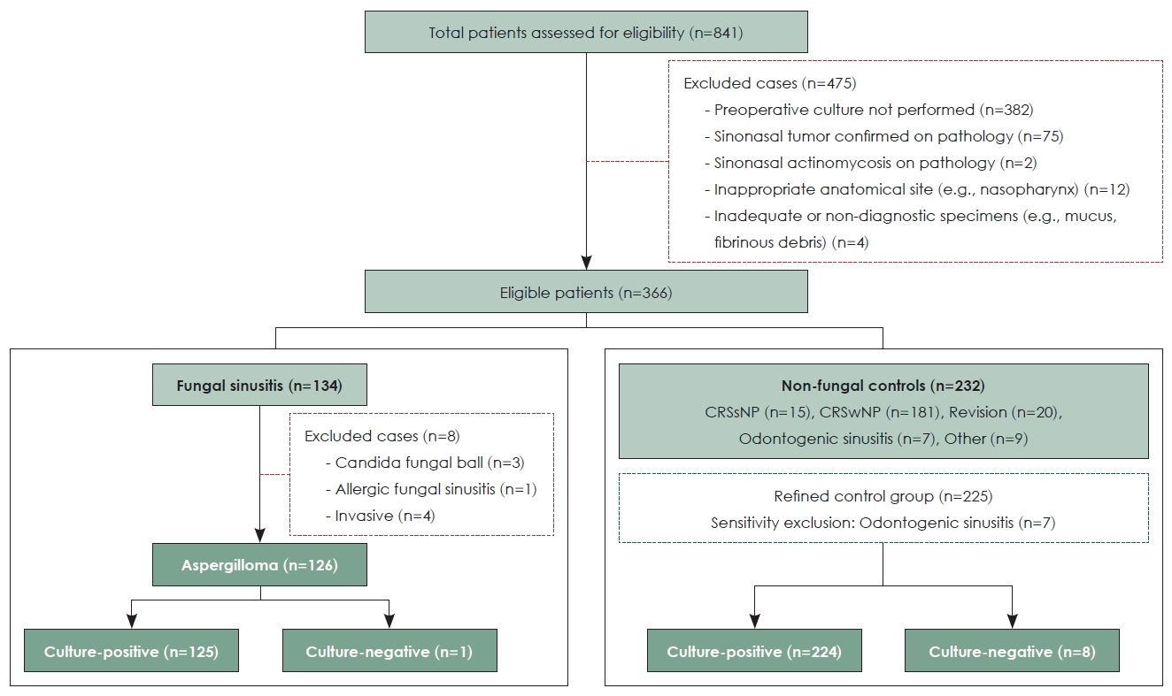
Flowchart of the patient selection and allocation process. CRSsNP, chronic rhinosinusitis without nasal polyps; CRSwNP, chronic rhinosinusitis with nasal polyps.
The control group was further stratified according to specific etiologies that could influence microbiological patterns: CRS without nasal polyps (CRSsNP) in 15 patients, of whom 13 had unilateral lesions; CRS with nasal polyps (CRSwNP) in 181 patients; CRS requiring revision surgery in 20 patients; odontogenic sinusitis in 7 patients; and other obstructive or inflammatory conditions in 9 patients.
Fungal sinusitis was classified according to its pathological subtype as fungus ball, allergic fungal sinusitis, or invasive fungal sinusitis [7]. To ensure microbiological and clinical homogeneity of the study population, only the predominant subgroup of patients with aspergilloma (n=126) was included in the primary analysis. A total of 8 patients were excluded from this analysis, comprising 3 patients with Candida fungal balls, 1 patient with allergic fungal sinusitis, and 4 patients with invasive fungal sinusitis [8,9]. The final aspergilloma group consisted of 125 culture-positive patients and 1 culture-negative patient.
To control for the potential confounding influence of conditions with distinctive microbiological characteristics, such as odontogenic sinusitis, a separate sensitivity analysis was performed after excluding patients with odontogenic sinusitis from the control group.
This study was approved by the Institutional Review Board of Kangdong Sacred Heart Hospital, and the requirement for written informed consent was waived (KANGDONG IRB 2026-02-004).
Diagnostic criteria for Aspergilloma
Aspergilloma was confirmed when fungal hyphae were identified on pathological examination of surgically obtained specimens. Gomori methenamine silver and periodic acid-Schiff staining were performed to assess the morphological features characteristic of Aspergillus, including septate hyphae with 45-degree branching [10].
Sinonasal bacterial culture
In all patients, purulent secretions were collected from the middle meatus with a swab under nasal endoscopic guidance and submitted for bacterial culture. Preoperative cultures were obtained in the outpatient setting in 69 patients, comprising 40 patients in the aspergilloma group and 29 patients in the control group. For intraoperative cultures, only secretions retained in the middle meatus were sampled at the beginning of surgery and before any manipulation of the sinonasal mucosa; specimens were not obtained after opening the sinus or from internal fungal debris. This approach was adopted to maintain anatomical comparability with cultures obtained in the outpatient setting. Specimens were immediately transferred to the hospital microbiology laboratory, where organisms were identified using standardized culture methods [11]. Culture results were reviewed retrospectively from the electronic medical records.
Classification of bacterial culture results
Cultured organisms were classified hierarchically according to their microbiological characteristics. When a single organism was detected, it was classified as either a gram-positive or a gram-negative bacterium according to its gram-staining characteristics. Polymicrobial infection was defined as the detection of two or more organisms and was further subdivided as follows: when all detected organisms shared the same gram-staining characteristics, the culture result was classified into the corresponding gram category; when both grampositive and gram-negative bacteria were detected, the result was separately defined as a mixed infection. Cases reported only as normal flora, in which specific organisms could not be identified, were excluded from analyses based on bacterial characteristics. Cases in which no organism grew on culture were separately classified as culture-negative and were included in the analysis.
Definition of PP
In this study, PP were defined as organisms reported to be clinically associated with sinonasal or respiratory tract infections. This definition was based on evidence from previous studies identifying these organisms as pathogenic, irrespective of whether a given organism is considered part of the normal flora [5,12]. The following organisms were included in this category: Pseudomonas aeruginosa; Enterobacterales (including Enterobacter spp., Klebsiella spp., Citrobacter spp., Escherichia coli, Proteus spp., Serratia spp., and Morganella spp.), Acinetobacter spp., Stenotrophomonas spp., Achromobacter spp., Burkholderia spp., Haemophilus influenzae; and Aggregatibacter spp [13].
Clinical data collection
Patient age, sex, diagnosis, bacterial culture results, and pathological findings were collected retrospectively from the medical records. All personally identifiable information was removed during data collection, and the data were anonymized by assigning a unique identification number to each patient before analysis.
Statistical analysis
Statistical analyses were performed using R version 4.5.2 (R Foundation for Statistical Computing), with statistical significance defined as p<0.05. Categorical variables were presented as frequencies and percentages [n (%)], and continuous variables as the mean±SD. Categorical variables were compared between groups using the chi-square test or Fisher’s exact test, and continuous variables using the independentsamples t-test. Binary logistic regression analysis was performed with the detection of PP as the dependent variable and study group, age, sex, polymicrobial infection, and mixed infection as independent variables; crude odds ratios (cORs), adjusted ORs (aORs), and their 95% confidence intervals (CIs) were calculated. The diagnostic performance of the culture indicators, namely PP and gram-negative bacteria, for predicting aspergilloma was evaluated using sensitivity, specificity, positive predictive value, negative predictive value, and the area under the curve (AUC), and AUCs were compared using the DeLong test. Additional sensitivity analyses were performed after excluding cases reported as normal flora, after including only culture-positive cases, and after excluding patients with odontogenic sinusitis. A subgroup analysis restricted to preoperative cultures obtained in the outpatient setting was also performed.
Results
Clinical characteristics of the study population
A total of 358 patients were included in the analysis, comprising 126 patients in the aspergilloma group and 232 patients in the control group. The aspergilloma group consisted of 49 male (38.9%) and 77 female (61.1%), with a mean age of 68.33±11.54 years, whereas the control group consisted of 173 male (74.6%) and 59 female (25.4%), with a mean age of 50.26±16.25 years.
When the bacterial culture results were classified as a single gram-negative isolate, a single gram-positive isolate, polymicrobial infection, or normal flora, the aspergilloma group showed a higher proportion of women, an older mean age, and a significantly higher PP detection rate than the control group (p<0.001) (Table 1). As aspergilloma accounted for the majority of the fungal sinusitis subgroups, comprising 126 patients (94.0%), the analysis focused on the clinical characteristics and bacterial culture profiles of this group.
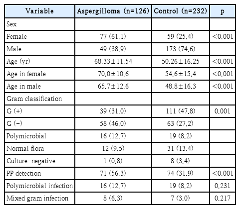
Clinical and demographic characteristics by group (aspergilloma vs. control)
Among the 232 patients in the control group, CRSwNP was the most common diagnosis, accounting for 181 patients (78.0%), followed by CRS requiring revision surgery in 20 patients (8.6%), CRSsNP in 15 patients (6.5%), of whom 13 had unilateral lesions, other obstructive or inflammatory conditions in 9 patients (3.9%), and odontogenic sinusitis in 7 patients (3.0%).
Overall distribution of sinonasal bacterial culture results
Bacterial culture patterns differed markedly between the aspergilloma and control groups. A single gram-negative isolate was identified in 46.0% (58/126) of the aspergilloma group, compared with 27.2% (63/232) of the control group, whereas a single gram-positive isolate was more common in the control group than in the aspergilloma group, at 47.8% and 31.0%, respectively.
Polymicrobial infection was identified in 12.7% (16/126) of the aspergilloma group and 8.2% (19/232) of the control group, and mixed infection involving both gram-positive and gram-negative bacteria accounted for a higher proportion in the aspergilloma group. Culture-negative results were observed in 1 patient (0.8%) in the aspergilloma group and 8 patients (3.4%) in the control group (Table 1).
Among the 19 control patients with mixed infection, only 1 (5.3%) had odontogenic sinusitis, which may exhibit distinctive microbiological characteristics, whereas the majority, comprising 15 patients (78.9%), belonged to the CRSwNP group. A sensitivity analysis was therefore performed after excluding the 7 patients with odontogenic sinusitis from the control group. No statistically significant differences in the overall culture-negative rate or in the frequencies of the major identified organisms were observed before and after exclusion (p>0.05).
Detection patterns of PP
The PP detection rate was significantly higher in the aspergilloma group than in the control group, at 56.3% (71/126) and 31.9% (74/232), respectively (p<0.001). The most commonly detected PP were gram-negative organisms, namely Pseudomonas aeruginosa in 17 patients (13.5%) and Enterobacterales, including Enterobacter in 20 patients (15.9%) and Klebsiella in 19 patients (15.1%) (Table 2). In the aspergilloma group, patients with PP detection predominantly harbored a single gram-negative isolate or a polymicrobial infection, and polymicrobial infection was significantly more frequent among patients with PP detection than among those without, at 18.3% (13/71) and 5.5% (3/55), respectively.
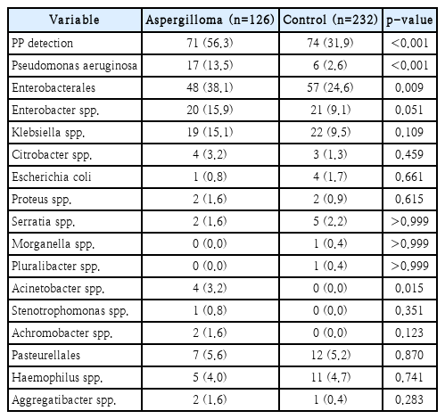
Patterns of PP by group (aspergilloma vs. control)
Microbial combinations according to culture type
Among the polymicrobial infections in the aspergilloma group, the combination of gram-negative and gram-positive bacteria was the most common pattern, accounting for 50.0% (8/16). Mixed infections most frequently involved Klebsiella spp. (43.8%, 7/16) or Pseudomonas aeruginosa (37.5%, 6/16). Three or more organisms were simultaneously detected in 3 patients, all of whom harbored gram-negative bacteria (Table 3).
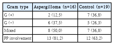
Microbial combinations in polymicrobial infection
Risk factors associated with potential pathogen detection
In the univariable analysis, PP detection was significantly associated with age (cOR 1.02, p=0.007), polymicrobial infection (cOR 4.23, p<0.001), and mixed infection (cOR 22.66, p=0.003).
In the multivariable logistic regression analysis, after adjustment for age, sex, and polymicrobial infection, patients in the aspergilloma group had an approximately 2.63-fold higher risk of PP detection than those in the control group (aOR 2.63; 95% CI, 1.51-4.58; p<0.001). Mixed infection was also identified as an independent predictor of PP detection (aOR 13.35; 95% CI, 1.39-128.04; p=0.025). In contrast, age, which was significant in the univariable analysis, lost significance in the multivariable analysis (aOR 1.01; 95% CI, 0.99-1.03; p=0.254), indicating that age was a confounding factor between the groups (Table 4).
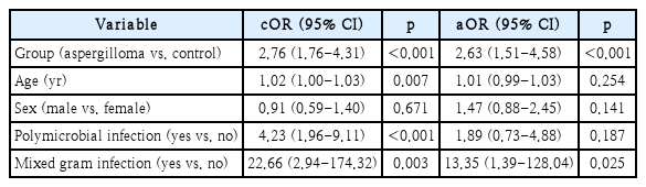
Logistic regression for factors associated with PP detection (n=358)
In the subgroup analysis restricted to cultures obtained in the preoperative outpatient setting, the diagnostic performance of PP detection in distinguishing the aspergilloma group from the control group remained similar to that observed in the overall analysis (AUC 0.625 vs. 0.622).
Diagnostic performance of culture-based predictors
To evaluate the value of bacterial culture results as preoperative diagnostic indicators, diagnostic performance was assessed in the overall cohort and in the subgroup restricted to cultures obtained in the preoperative outpatient setting (Table 5).

Diagnostic performance of culture predictors for aspergilloma
In the overall cohort, comprising 126 patients in the aspergilloma group and 232 patients in the control group, detection of gram-negative bacteria yielded a sensitivity of 57.1% (95% CI, 48.0-65.9), a specificity of 67.7% (95% CI, 61.2-73.6), and an AUC of 0.624 (95% CI, 0.571-0.677).
To exclude potential distortion arising from intraoperative sampling and to reflect performance as a preoperative marker, only cultures obtained in the preoperative outpatient setting were analyzed. This subgroup comprised 40 patients in the aspergilloma group and 29 patients in the control group. The specificity and positive predictive value of gram-negative bacterial detection increased to 72.4% (95% CI, 52.8-87.3) and 73.3% (95% CI, 54.1-87.7), respectively, and the AUC increased slightly to 0.637 (95% CI, 0.523-0.751).
The model combining gram-negative bacteria, age, and sex demonstrated high diagnostic performance, with an AUC of 0.858 (95% CI, 0.818-0.898). However, compared with the model containing only age and sex (AUC 0.849), the incremental diagnostic value of gram-negative bacterial detection was not statistically significant (DeLong p=0.240).
Discussion
In this study, we analyzed preoperative sinonasal bacterial culture patterns in patients with pathologically confirmed aspergilloma who underwent endoscopic sinus surgery, and we identified high detection rates of both PP, as defined in this study, and gram-negative bacteria as significant microbiological features of aspergilloma. These findings are consistent with recent comparative culture-based research and microbiome studies [14]. Bacterial culture results should nonetheless be interpreted as an adjunctive screening tool that supports clinical judgment rather than as a single independent indicator for the definitive diagnosis of aspergilloma. Even so, our findings suggest that bacterial culture may serve not only as a tool for antibiotic selection but also as an adjunctive screening indicator that raises clinical suspicion of aspergilloma requiring surgical treatment.
Among the 358 patients included in the analysis, the aspergilloma group had an older mean age, a higher proportion of women, and a significantly higher potential pathogen detection rate than the control group (p<0.001). These demographic patterns were similar to those reported in previous studies of fungal balls [15]. The proportion of single gram-negative isolates was also significantly higher in the aspergilloma group than in the control group, consistent with culture-based comparative studies reporting a predominance of gram-negative bacteria in noninvasive fungal sinusitis [16,17].
In the multivariable logistic regression analysis, aspergilloma remained independently associated with potential pathogen detection (aOR 2.63; 95% CI, 1.51-4.58; p<0.001), and mixed infection was also strongly associated with potential pathogen detection (aOR 13.35; 95% CI, 1.39-128.04; p=0.025). These associations remained consistent in sensitivity analyses that excluded patients whose culture results were reported only as normal flora and patients with odontogenic sinusitis (Table 6), supporting an association between potential pathogen detection and the microbiological environment of aspergilloma, regardless of whether normal flora was reported or patients with odontogenic sinusitis were included.

Robustness of the group–PP association across sensitivity analyses
Although surgical removal is the primary treatment for aspergilloma and is critical for achieving cure and favorable outcomes, clinical symptoms and imaging findings, including calcification, may be inconclusive in the early stage, and surgical treatment may therefore be delayed while patients receive repeated courses of antibiotics. Despite these diagnostic challenges, the present findings indicate that sinonasal bacterial culture results may provide an adjunctive clue that raises suspicion of aspergilloma. In particular, the detection of PP or gram-negative bacteria in cultures obtained during outpatient evaluation or from the middle meatus according to our institutional standard protocol may help clinicians consider the possibility of aspergilloma before definitive radiological signs become apparent, thereby supporting earlier consideration of surgical management.
The organisms predominantly detected in the aspergilloma group included Pseudomonas aeruginosa, Enterobacterales such as Enterobacter, Klebsiella, and Citrobacter, and Acinetobacter spp., all of which are pathogenic organisms not considered part of the normal sinonasal flora. Similar bacterial distributions have also been reported in microbiome studies of fungal balls and CRS [18].
Differences in bacterial community composition between patients with aspergilloma and those with nonfungal CRS have also been demonstrated in studies based on 16S rRNA sequencing [19], and the present findings indicate that these differences may be reproduced using culture-based analysis. To avoid statistical selection bias, culture-negative patients were not excluded from the primary analysis, an approach intended to objectively reflect the culture-positive rate observed in clinical practice.
This predominance of specific organisms may reflect microbiota dysbiosis resulting from chronic inflammation and repeated antibiotic exposure, and it suggests that fungal-bacterial interactions may contribute to the pathophysiology [6]. In particular, the microbiological differences observed between the aspergilloma and control groups demonstrate that bacterial culture results may serve as an adjunctive marker for differentiating the two conditions.
The predominance of gram-negative bacteria in the aspergilloma group may also help create a microenvironment favorable for fungal colonization by weakening mucosal defense mechanisms and promoting biofilm formation. Interactions between Aspergillus and bacteria and their potential involvement in biofilm formation have been reported in several clinical studies [20].
The independent associations of polymicrobial and mixed infections with potential pathogen detection suggest that aspergilloma may be associated with alterations in a complex microbial community rather than with infection by a single pathogen [21]. Mixed infections involving both gram-positive and gram-negative bacteria accounted for 50% of polymicrobial infections, with Klebsiella spp. and Pseudomonas aeruginosa being the organisms most commonly involved. These findings suggest that interactions among specific pathogenic organisms may play an important role in Aspergillus colonization.
Age was significantly associated with potential pathogen detection in the univariable analysis (cOR 1.02, p=0.007), but was no longer significant in the multivariable model that included age, sex, and polymicrobial infection (aOR 1.01, p=0.254). This finding indicates that age acted as a confounding factor in the between-group comparison and supports the interpretation that aspergilloma itself is a major independent factor associated with potential pathogen detection. A similar pattern has been reported in previous studies of fungal balls [14].
Notably, the direction and statistical significance of the main findings remained consistent in sensitivity analyses that either included or excluded patients whose cultures yielded only normal flora. This finding suggests that the association between potential pathogen detection and aspergilloma was not substantially affected by the presence of normal flora and that microbiota dysbiosis itself may represent an important pathophysiological signal. The observed organism distribution was similar to that reported in previous large-scale studies of sinonasal bacterial cultures [22]. Statistical significance also remained unchanged in a sensitivity analysis excluding the 7 patients with odontogenic sinusitis, a condition with distinctive microbiological characteristics, from the control group. These findings indicate that a specific disease subgroup within the control group did not distort the overall microbiological pattern and support the reliability of the study conclusions.
The most important clinical implication of this study is that the detection of PP or gram-negative bacteria in sinonasal bacterial cultures may provide adjunctive evidence for reconsidering repeated antibiotic treatment and for pursuing further radiological evaluation or a transition to surgical treatment in light of the possibility of aspergilloma. Culture results may assist clinical decision-making during the initial stage of medical treatment or in the outpatient setting before imaging examinations such as CT, potentially helping to reduce unnecessary long-term antibiotic use. Bacterial culture also has the clinical advantages of being substantially less expensive than CT, posing a lower economic burden, and being readily performed in the outpatient setting without complex preparation. Therefore, for patients for whom immediate CT is not feasible because of cost, limited access to equipment, or restricted CT availability in certain countries or primary care settings, bacterial culture may have substantial value as an economical and efficient initial screening tool, and it may complement radiological examinations and improve clinical efficiency.
Additional analyses of the diagnostic performance of preoperative bacterial culture results showed that the AUCs of the individual culture indicators ranged from 0.593 to 0.637, which limits their interpretation as stand-alone diagnostic indicators. However, when only preoperative outpatient culture data were analyzed, the specificities and positive predictive values of potential pathogen detection and gram-negative bacterial detection were 72.4% or higher, which is clinically noteworthy. Therefore, rather than being used as stand-alone diagnostic criteria, the high detection rate of gram-negative bacteria should be interpreted as a characteristic microbiological pattern observed more frequently in the aspergilloma group.
This study has several limitations. First, its retrospective, single-center design introduces the possibility of selection bias inherent in a surgical cohort. Patients undergoing surgical treatment are often more symptomatic or refractory than those treated in primary care settings, which may limit the generalizability of the findings. Second, although culturenegative patients were retained as a separate category in the primary analysis to minimize statistical distortion, validation in a larger prospective study is needed. Third, the timing of specimen collection varied between the preoperative outpatient clinic and the operating room. Although intraoperative specimens were strictly limited to secretions from the middle meatus obtained before sinus opening, in accordance with our institutional standard surgical protocol to ensure anatomical comparability, subtle confounding related to differences in collection timing and in the duration of prior antibiotic exposure cannot be completely excluded. Fourth, because this study was based on conventional bacterial culture, it had technical limitations in fully capturing difficult-to-culture anaerobic organisms and dynamic changes in the overall microbiome that can be assessed using next-generation sequencing. Finally, the microbiological characteristics identified in this study may not be representative of clinically distinct subtypes such as allergic fungal rhinosinusitis or invasive fungal sinusitis and therefore cannot be broadly generalized to all fungal sinusitis subtypes. Nevertheless, this study has substantial clinical value, as it provides specific statistical measures of diagnostic performance and multiple sensitivity analyses of the screening utility of bacterial culture in aspergilloma, the most common form encountered in clinical practice.
Supplementary Materials
Korean translation of this article is available with the Online-only Data Supplement at https://doi.org/10.3342/kjorl-hns.2026.00416.
Notes
Acknowledgments
None
Author Contribution
Conceptualization: Kyubo Kim. Data curation: Ji Eun Kim, Ji Young Lim. Formal analysis: Ji Eun Kim. Investigation: Ji Eun Kim, Ji Young Lim. Methodology: Kyubo Kim. Supervision: Kyubo Kim. Writing—original draft: Ji Eun Kim. Writing—review & editing: Ji Eun Kim, Kyubo Kim.