How I Do It: The Submental Flap in Head and Neck Reconstruction
악하피판을 이용한 두경부 재건
Article information

Abstract
The submental flap is a reliable and versatile regional reconstructive option for small-to-moderate defects of the oral cavity, buccal mucosa, oropharynx, hypopharynx and lower face. Based on the submental artery, a consistent branch of the facial artery, this pedicled flap offers thin, pliable tissue with excellent color and texture match to the surrounding facial skin. Compared to microvascular free tissue transfer, it provides shorter operative time, lower perioperative risk, and ease of harvest, making it particularly advantageous in elderly patients and those with significant comorbidities. In this article, I describe my current technique for harvesting and applying the submental flap, refined over years of experience. Key principles include mandatory inclusion of the ipsilateral anterior belly of the digastric muscle for perforator preservation, contralateral-to-ipsilateral subplatysmal dissection, meticulous identification of venous outflow pathways during concurrent neck dissection, and careful management of oncologic risk in oral cavity cancer. With proper planning and technique, the submental flap provides consistent, reproducible outcomes with minimal donor-site morbidity.
Introduction
The submental flap―originally described by Martin, et al. [1] in 1993―has become my first-line regional reconstructive option for small-to-moderate head and neck defects. Its reliable vascular anatomy based on the submental artery, thin and pliable skin paddle with excellent facial color match, and a well-concealed donor-site scar in the submental crease make it particularly suitable for reconstruction of the oral cavity, oropharynx, and lower face [2,3].
Over my clinical experience, I have refined a consistent approach that prioritizes perforator preservation through the anterior belly of the digastric muscle, reliable venous outflow identification during concurrent neck dissection, and individualized patient selection based on anatomical and oncologic factors. In this article, I describe my current technique and share the key technical pearls that have shaped my practice.
Indications and Patient Selection
The submental flap is best suited for small-to-moderate defects of the oral cavity, floor of mouth, buccal mucosa, oropharynx, hypopharynx and lower facial skin. Its pliability and favorable color match make it especially advantageous for reconstruction of mobile mucosal surfaces and visible cutaneous regions.
In my practice, the submental flap is most beneficial in the following patient groups:
- Elderly patients (generally ≥70 years) with multiple comorbidities
- Patients who are suboptimal candidates for lengthy microvascular procedures
- Cases where operative time minimization is clinically important
- Patients desiring faster postoperative recovery
I apply the following contraindications in my clinical decision-making (Table 1).
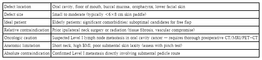
Indications and contraindications for the submental flap
A critical oncologic consideration in oral cavity cancer is the theoretical risk of inadvertently transposing Level I lymph nodes to the recipient bed. In my practice, I address this with thorough preoperative imaging and meticulous perivascular dissection during concurrent neck dissection to ensure complete lymph node clearance—an approach supported by published clinical experience [4].
Preoperative Planning and Flap Design
Patient assessment
Preoperative planning begins with systematic evaluation of neck anatomy, skin laxity, and vascular accessibility. The pinch test—grasping the submental skin between the thumb and forefinger—remains the most reliable bedside method to estimate the available vertical flap height. In my experience, 6-8 cm of vertical skin paddle can typically be harvested without undue tension on primary closure. Anatomic factors that may preclude flap use—short neck morphology, high BMI, or prior submental scarring—should be identified at this stage.
Flap design
The flap is designed in an elliptical shape centered on the submental area, approximately 1 cm below the mandibular border to ensure the resultant scar is concealed within the submental crease (Fig. 1). Key design principles include:
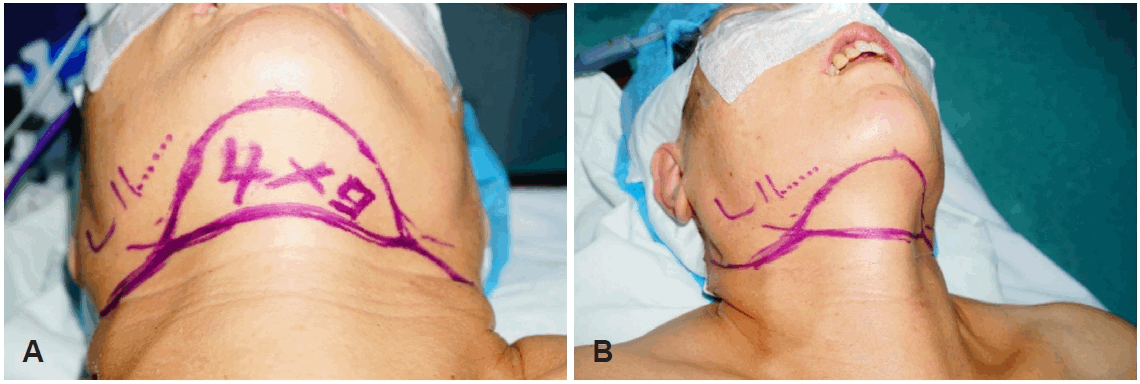
Design of the submental island flap. The elliptical skin paddle is outlined over the submental region with its long axis oriented along the lower border of the mandible; the planned dimensions (width×length, cm) are indicated within each marking. A: A 4×9 cm skin paddle with the planned submandibular and neck-dissection incisions. B: A 5×8 cm skin paddle.
- Superior border: 1 cm below the inferior mandibular border
- Vertical height: determined by the pinch test (typically 6-8 cm)
- Horizontal width: tailored to defect dimensions; can extend bilaterally toward both mandibular angles if a larger paddle is required
- Pedicle side: ipsilateral to the dominant facial artery; confirmed by Doppler if needed
The submental artery—diameter approximately 1.0-1.7 mm, pedicle length 50-80 mm—originates from the facial artery medial to the submandibular gland and courses along the mylohyoid muscle, giving off perforating branches through the anterior belly of the digastric muscle to the overlying skin [5,6]. This anatomy determines the critical technical steps described below.
Venous drainage anatomy
In contrast to the arterial supply, which arises consistently from the facial artery, the venous outflow of the submental flap is anatomically variable and warrants explicit preoperative and intraoperative attention. The submental vein accompanies the submental artery and, in most patients, drains through the facial vein and common facial vein into the internal jugular vein. It does not, however, invariably follow this route. In a clinical series of 70 submental flap harvests, venous return was to the internal jugular vein in 72.9% of cases but to the external jugular vein in the remaining 27.1% [7]. A separate multicenter series of 83 oral cancer reconstructions similarly reported venous drainage to the internal jugular vein in 68.7%, the external jugular vein in 21.7%, and the anterior jugular vein in 9.6% of cases [8]. The facial vein is therefore the dominant—but by no means the only—route of venous outflow from the flap.
This variability has direct surgical implications. Because the submental vein may return through the external jugular or anterior jugular system rather than the facial vein, preservation of the common facial vein alone does not guarantee adequate venous drainage of the flap. In every case, the dominant draining vein should be positively identified during elevation and deliberately protected throughout any concurrent neck dissection. Inadvertent ligation of an unrecognized external or anterior jugular outflow channel during Level I-II clearance is a readily avoidable cause of venous congestion and flap loss.
Surgical Technique
Step 1: patient positioning
The patient is placed supine with a shoulder roll to achieve neck extension. The head is stabilized on a donut headrest. Importantly, the shoulder roll is removed prior to donor-site closure to reduce tension on the primary closure line.
Step 2: flap marking and skin incision
The previously designed elliptical outline is confirmed with the neck in neutral position. The superior incision is placed 1 cm below the mandibular border. Skin incision is carried through the dermis, subcutaneous tissue, and platysma muscle (Fig. 2).
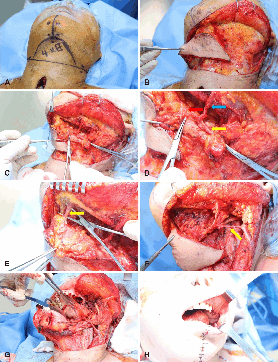
Step-by-step harvest of the submental island flap. A: Flap design with the skin paddle marked over the submental region. B: Subplatysmal elevation of the flap proceeding from the contralateral side. C: Identification of the pedicle vessels during concurrent neck dissection. D: Exposure of the facial artery and submental artery (blue arrow: distal part of facial artery, red arrow: submental artery, yellow arrow: proximal part of facial artery). E: Ligation of the distal part of submental artery (yellow arrow: distal submental artery). F: Completed neck dissection with the prepared flap including ipsilateral anterior belly of digastric muscle; the venous drainage to the external jugular vein is identified (yellow arrow: vein drainage to external jugular vein). G: Intraoral insetting of the flap. H: Completion of surgery.
Step 3: subplatysmal dissection—contralateral to ipsilateral direction
This step is the most critical in my technique. Dissection proceeds in the subplatysmal plane from the contralateral to the ipsilateral side. I begin with the contralateral submental vessels, ligating them first, then elevate the skin paddle from the distal (contralateral) to the proximal (ipsilateral, pedicle) direction. On the contralateral side, dissection proceeds superficial to the anterior belly of the digastric muscle; on the ipsilateral side, it proceeds deep to the digastric muscle to preserve the submental artery perforators.
This directional approach offers two advantages: 1) it allows early control of the contralateral submental vessels, and 2) it protects the perforating branches of the submental artery on the ipsilateral side throughout the elevation.
Step 4: identification of the marginal mandibular nerve, facial vessels, and integrated level I dissection
Dissection continues to the ipsilateral submandibular region. The marginal mandibular nerve is identified early at the mandibular notch and carefully preserved throughout. The facial artery and vein are identified superior to the submandibular gland. The distal facial artery (superior to the gland) is ligated to allow pedicle mobilization; however, the venous outflow must be preserved with particular care. Although the submental vein most often drains through the common facial vein into the internal jugular vein, it returns via the external jugular or anterior jugular vein in a substantial minority of patients; the dominant draining vein should therefore be identified and preserved in each individual case, rather than assuming the common facial vein to be the sole outflow.
Level I nodal dissection is integrated into this step rather than being performed as a separate procedure. With the nerve preserved and the vessels localized, dissection is carried proximal to the facial vessels and the perifacial nodes are removed in continuity as the pedicle is exposed. Identifying the vessels before dividing any perifacial tissue is essential, as it prevents inadvertent injury to the pedicle during node clearance. If gross nodal disease is encountered at Level I during this dissection, the submental flap is abandoned and reconstruction is converted to a free flap.
Step 5: submandibular gland mobilization and pedicle dissection
The submandibular gland is separated from the submental artery as it branches anteriorly. The gland may be excised or mobilized to improve arc of rotation if needed. As the flap is freed from the mandibular margin, the distal (terminal) branch of the submental artery—which continues over the inferior border of the mandible toward the chin and lower lip—is ligated and divided (Fig. 2E). Pedicle dissection is then continued proximally along the facial artery toward its origin to maximize pedicle length.
Step 6: inclusion of the anterior belly of the digastric muscle
With the pedicle dissected, the ipsilateral anterior belly of the digastric muscle is detached from its hyoid and mandibular attachments and carried with the flap. Because the submental artery courses medial to this muscle in approximately 70%-80% of cases and gives off perforating branches to the overlying skin, its inclusion is essential for reliable perforator preservation [6]. In my experience, inclusion of the mylohyoid muscle is not routinely necessary—the digastric anterior belly alone provides sufficient perforator protection.
Step 7: flap transfer and inset
Once elevation is complete, the flap is transferred to the recipient site. For intraoral reconstruction, a transmylohyoid tunnel is created to pass the flap without external skin bridging. The pedicle is positioned without tension, torsion, or kinking, and the flap is inset with absorbable sutures. A closed-suction drain is placed at the donor site. Primary closure of the donor site is typically achievable; the shoulder roll is removed at this point to reduce tension.
Postoperative Management
Postoperative monitoring focuses on early detection of vascular compromise, which most commonly occurs within the first 48 hours. The skin paddle is assessed clinically for color, turgor, and capillary refill; portable Doppler examination of the submental pedicle is performed at regular intervals.
Standard postoperative protocols in my practice include:
- Head elevation at 30° to reduce venous congestion
- Neutral or slightly flexed neck position to avoid donorsite tension
- Closed-suction drain removal after 3-5 days, once output is minimal
- Suture removal on postoperative days 7-10
- Soft or liquid diet after postoperative 7 days for intraoral flaps
Modifications and Extended Applications
Several modifications have been described to extend the reach or augment the perfusion of the submental flap. In the reverse-flow submental flap, the proximal facial artery is ligated and the flap is perfused by retrograde flow from the distal branches, extending the arc of rotation toward the upper third of the face and the periorbital region [9]. A hybrid technique has also been proposed, in which the flap is raised as a pedicled flap but its venous outflow is augmented by microvascular anastomosis to a recipient vein to address marginal pedicle length or venous congestion. Pedicle length has likewise been extended by ligating the common facial vein and relying on reverse flow through the retromandibular vein into the external jugular vein, reportedly adding up to 5 cm of venous pedicle [10].
In my own practice, however, I am cautious about routinely adopting these modifications. Each adds technical complexity, and reverse-flow perfusion in particular introduces a degree of unpredictability in flap viability. More fundamentally, the principal advantages of the submental flap—its simplicity, speed, and avoidance of microsurgery—are largely lost once a microvascular venous anastomosis or a reverseflow maneuver becomes necessary. In my view, when a defect genuinely exceeds the reliable reach or perfusion of a standard pedicled submental flap, it is safer and more reproducible to proceed with a well-established free flap such as the radial forearm free flap, which provides predictable, robust antegrade perfusion. Put simply, if the reconstruction is going to demand a microsurgical commitment or depend on retrograde flow, a free flap is usually the more dependable choice than a modified submental flap.
Results
In my clinical experience, the submental flap has been applied to a range of head and neck defects including oral cavity, oropharynx, hypopharynx and lower facial reconstruction. The majority of patients were elderly with significant medical comorbidities who were not candidates for prolonged free flap procedures.
Flap survival rates in my series have been consistently high, with most partial or complete losses attributable to venous congestion in cases where concurrent Level I neck dissection inadvertently compromised venous outflow—reinforcing the critical importance of identifying and preserving the flap’s actual venous outflow pathway—whether through the facial, external jugular, or anterior jugular vein. Donorsite morbidity was minimal in all cases, and the concealed submental scar provided consistently satisfactory cosmetic outcomes.
Published comparative data support the oncologic equivalence of the submental flap to radial forearm free flap in appropriately selected patients (Table 2). Sittitrai, et al. [11] demonstrated comparable locoregional control and 5-year survival in a propensity-matched cohort of oral cavity cancer patients, while also reporting lower donor/recipient complication rates, shorter hospital stay, and reduced cost with the submental approach.
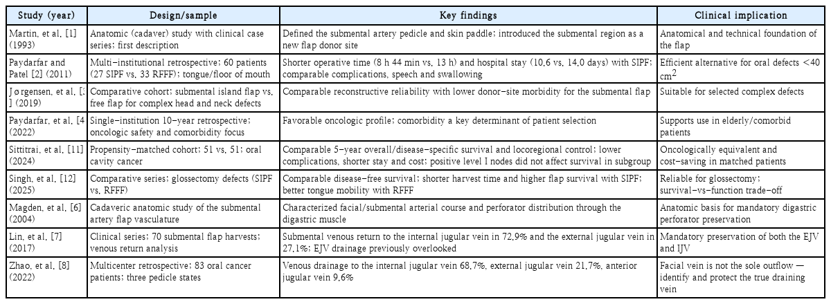
Summary of key studies on the submental flap
Discussion
The submental flap occupies a distinct and valuable niche in the head and neck reconstructive armamentarium. Unlike the radial forearm free flap—which remains the gold standard for oral cavity reconstruction—the submental flap does not require a two-team approach, microsurgical infrastructure, or prolonged operating time. These practical advantages are particularly meaningful in resource-limited settings and in high-risk patients for whom operative time directly correlates with perioperative morbidity.
The oncologic safety of the submental flap in oral cavity cancer has been a point of ongoing debate since its introduction. Early concerns centered on the theoretical risk of transposing Level I lymph nodes—which lie in the proximity of the submental pedicle—to the recipient bed. However, multiple series have now demonstrated that with meticulous dissection and thorough preoperative imaging, the submental flap can be used safely even in the setting of concurrent Level I-II neck dissection [4,11].
The most common cause of flap failure in my experience has been venous congestion secondary to compromise of the venous outflow during neck dissection. This underscores a key technical principle that distinguishes experienced from novice submental flap surgeons: venous outflow management is as critical as arterial inflow. Crucially, the facial vein is not the only major outflow channel: published series have shown that the submental vein drains into the external jugular vein in roughly 21%-27% and into the anterior jugular vein in approximately 10% of patients, rather than into the common facial vein and internal jugular vein [7,8]. Careful preoperative and intraoperative assessment of venous anatomy— and deliberate preservation of whichever vein (facial, external jugular, or anterior jugular) carries the dominant outflow in the individual patient—should therefore be standard practice.
Recent comparative studies have consistently confirmed the safety and efficacy of the submental flap. Singh, et al. [12] reported comparable disease-free survival between submental island pedicled flap and radial forearm free flap in glossectomy patients, while noting shorter harvest time and higher flap survival with the submental approach. These findings, combined with the demonstrated cost advantages [11], suggest that the submental flap may be underutilized in current practice.
Surgical tips
Tip 1. Always dissect from contralateral to ipsilateral direction
Proceeding from the distal (contralateral) end toward the pedicle allows early control of the contralateral submental vessels and protects the ipsilateral perforators throughout the dissection. This directional principle is the single most important technical detail in my technique.
Tip 2. Always include the ipsilateral anterior belly of the digastric muscle—but not the mylohyoid
The submental artery perforators pass through the anterior belly of the digastric muscle to reach the skin. Its inclusion is non-negotiable for reliable flap perfusion. The mylohyoid muscle does not need to be included in routine cases.
Tip 3. Identify and preserve the true venous outflow—the facial vein is not the only route
During concurrent neck dissection, the most frequent cause of venous congestion is inadvertent ligation of the flap’s draining vein. The facial vein is the dominant outflow in most patients, but it is not the only one: the submental vein drains via the external jugular vein in roughly one-quarter and via the anterior jugular vein in about one in ten patients [7,8]. Trace and confirm the actual venous drainage pathway—facial, external jugular, or anterior jugular—before ligating any vein in the Level I-II region, and preserve whichever channel carries the dominant outflow.
Tip 4. Recognize when a free flap is the safer choice
Reverse-flow and venous-augmentation maneuvers can extend the reach or drainage of the flap, but they add complexity and erode the predictability and microsurgery-free simplicity that make the submental flap attractive in the first place. When a defect exceeds the reliable reach or perfusion of a standard pedicled submental flap, a well-established free flap such as the radial forearm free flap is usually the safer and more reproducible choice.
Tip 5. Manage Level I oncologic risk proactively in oral cavity cancer
Preoperative CT, MRI, or PET-CT is mandatory to exclude Level I metastasis before using this flap in oral cavity cancer. During neck dissection, meticulous perivascular dissection ensures complete lymph node clearance around the pedicle, virtually eliminating the risk of inadvertent node transfer.
Tip 6. Create an adequate transmylohyoid tunnel for intraoral reconstruction
When passing the flap intraorally, the mylohyoid tunnel must be generously sized—sufficient to accommodate the flap and pedicle without compression. Inadequate tunnel diameter is a common cause of postoperative pedicle kinking and venous congestion. Verify there is no tension or torsion after positioning.
Tip 7. Remove the shoulder roll before donor-site closure
Donor-site closure should always be performed with the neck in a neutral or slightly flexed position. Closing under neck extension (shoulder roll in place) will produce excess tension on the closure line, increasing the risk of wound dehiscence or a widened scar.
Tip 8. Use the pinch test as the definitive guide for flap height
The vertical dimension of the flap must be determined by the pinch test in every case—not by the defect size alone. Overestimating available skin laxity results in donor-site closure under excessive tension. A conservative estimate of 6-8 cm is appropriate in most patients.
Conclusion
The submental flap remains a highly versatile and dependable option for head and neck reconstruction, particularly in patients who are poor candidates for microvascular free tissue transfer. By adhering to the key anatomical principles described here—mandatory inclusion of the anterior belly of the digastric muscle, contralateral-to-ipsilateral directional dissection, meticulous venous outflow preservation, and careful oncologic preparation in oral cavity cancer—consistent and successful outcomes can be achieved. With proper planning and technique, the submental flap represents an efficient, reproducible, and cosmetically superior reconstructive tool that deserves broader application in head and neck surgery.
Notes
Acknowledgments
This study was reviewed by the Institutional Review Board of Seoul St. Mary’s Hospital, The Catholic University of Korea, and was granted an exemption from review (IRB No. VC26ZASI0191).