Transplantation of Stem Cells to Modulate Spiral Ganglion Neuronal Density
줄기세포 이식을 통한 나선신경절 신경세포 밀도 조절
Article information

Abstract
Spiral ganglion neurons (SGNs) constitute the principal neural substrate for cochlear implantmediated auditory stimulation, and progressive SGN degeneration may limit the functional benefit of cochlear implantation. Stem-cell transplantation has therefore emerged as a potential strategy to preserve, replace, or increase the neuronal population within the spiral ganglion. Early studies demonstrated the survival and trophic effects of neural stem cells, followed by evidence of migration toward Rosenthal’s canal and modulation of SGN populations. Human mesenchymal stem cells and human embryonic stem-cell-derived otic progenitors subsequently provided the evidence of increased neuronal populations and functional auditory improvement. More recently, it has been shown that human dermal fibroblast-derived induced neural stem cells enhance cochlear neural structures, central auditory connectivity, and electrically evoke auditory responses. Collectively, these studies suggest that stem-cell therapy may improve the neural substrate for cochlear implantation not only through neuronal replacement but also through trophic preservation, targeted migration, and functional neural integration.
Introduction
Spiral ganglion neurons (SGNs) are the primary auditory neurons that transmit information from the cochlea to the central auditory pathway [1]. Following severe sensorineural hearing loss, SGNs may undergo progressive degeneration [2], particularly after loss of cochlear hair cells. Because cochlear implants bypass damaged hair cells and directly stimulate auditory neurons, preservation of an adequate SGN population is considered an important biological prerequisite for effective cochlear implantation.
The limited regenerative capacity of mammalian SGNs has stimulated considerable interest in cell-based regenerative strategies. Initial approaches focused on transplantation of neural stem cells to provide trophic support to the injured cochlea. Subsequent studies demonstrated that transplanted cells could migrate toward Rosenthal’s canal, where SGNs are located, and potentially contribute to or preserve the neuronal population. Human-derived mesenchymal stem cells (MSCs) subsequently provided evidence for increased SGN numbers, while human embryonic stem-cell-derived otic pro-genitors demonstrated a more lineage-directed approach and functional auditory improvement.
More recent work has shifted toward improving the specificity of transplanted cells and their localization within the injured cochlea [3]. Strategies have included chemotactic recruitment, alternative intracochlear delivery routes, lineagedirected otic progenitors, and directly converted human induced neural stem cells.
The purpose of this review is to summarize in vivo celltransplantation studies that directly address the SGN population or auditory neural function, with particular emphasis on cell type, transplantation route, migration, neuronal differentiation, changes in SGN-related structures, and functional outcomes. In vitro-only studies are not considered.
Cell Types Used for SGN-Directed Transplantation
The development of cell-based SGN regeneration can be viewed as a progression from relatively nonspecific neural stem cells toward increasingly lineage-directed and humanderived neural progenitors. The earliest studies used neural stem cells without specific auditory lineage specification. Iguchi, et al. [4] investigated neural stem-cell transplantation primarily as a means of providing trophic support to the injured inner ear. Parker, et al. [5] subsequently demonstrated that neural stem cells injected into the scala tympani could migrate throughout the cochlea, including toward the region of Rosenthal’s canal, and express markers associated with cochlear neural cells.
Lang, et al. [6] expanded the approach to embryonic stem cells in an ouabain-induced auditory neuropathy model, demonstrating the feasibility of transplantation of pluripotent cells into an injured auditory system.
The use of human cells represented an important subsequent step. Cho, et al. [7] transplanted neural-differentiated human MSCs into the scala tympani of guinea pigs with ouabain-induced auditory neuropathy and reported an increase in SGN number.
Chen, et al. [8] subsequently introduced human embryonic stem-cell-derived otic progenitors, representing a major transition toward developmental lineage specification. These cells were generated through directed differentiation toward an otic lineage before transplantation and subsequently demonstrated auditory neuronal characteristics in vivo.
The most recent development is represented by Chang, et al. [9], who generated induced neural stem cells directly from human dermal fibroblasts and transplanted these cells into the scala tympani of an auditory-neuropathy animal model (Table 1).
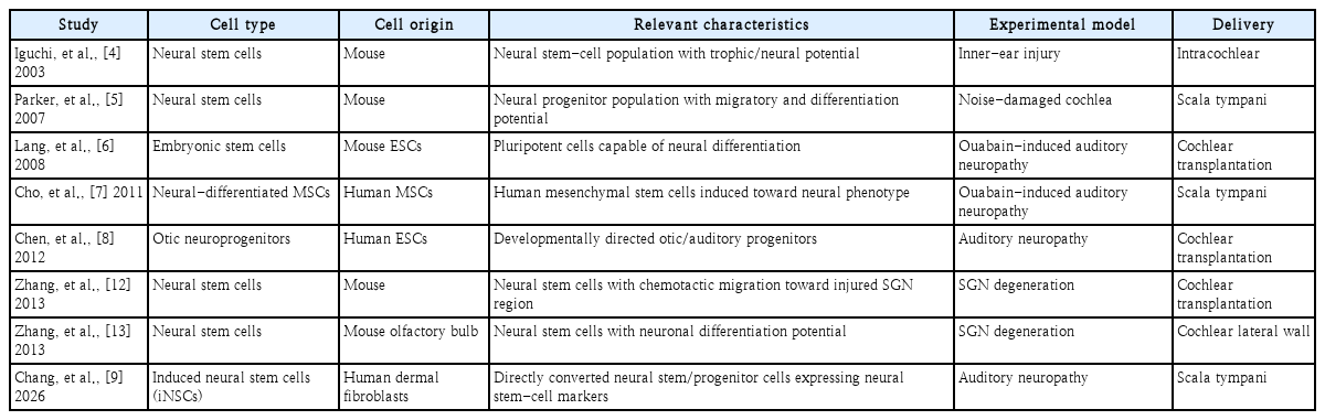
Cell types used for in vivo transplantation studies relevant to SGN regeneration
Biological Effects of Stem-Cell Transplantation
The biological effects of transplantation vary substantially among studies. Some primarily demonstrate survival and migration, whereas others demonstrate increased SGN populations, neuronal differentiation, or functional auditory improvement.
From Trophic Support to SGN Population Modulation
The earliest rationale for cell transplantation was not necessarily neuronal replacement. Instead, transplanted neural stem cells were viewed as biological sources of trophic factors capable of supporting injured endogenous neurons.
Iguchi, et al. [4] provided early evidence for this concept, demonstrating that neural stem-cell transplantation could provide trophic support to the injured inner ear. This approach is biologically relevant because SGN degeneration is often progressive. After the initial cochlear insult, a population of residual neurons may remain viable but vulnerable to secondary degeneration. A transplanted cell population capable of secreting neurotrophic molecules could therefore preserve neurons that otherwise would have been lost. However, trophic preservation alone cannot restore neurons that have already disappeared. This limitation stimulated interest in determining whether transplanted cells could actually enter the SGN compartment and increase the neuronal population.
Parker, et al. [5] provided important evidence in this regard. Neural stem cells injected into the scala tympani of noisedamaged animals migrated throughout the cochlea and were detected in the region of Rosenthal’s canal. The study also reported a small but significant increase in Type I SGN-associated cells following transplantation. The interpretation of increased SGN numbers requires caution. An increase in neuronal counts does not necessarily indicate direct replacement by transplanted cells. It may result from donor-cell differentiation, preservation of endogenous SGNs, trophic effects, or a combination of these mechanisms. Nevertheless, these findings established the important concept that trans planted cells can modulate the SGN compartment (Table 2).
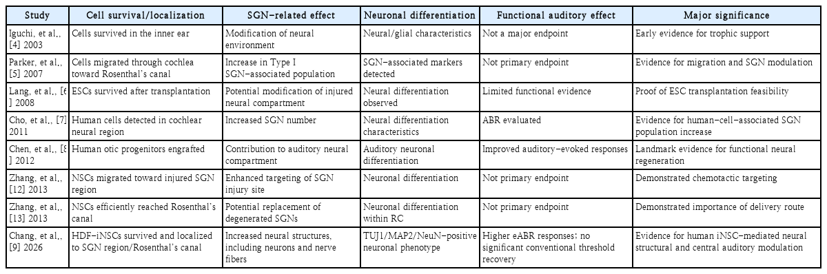
Effects of transplanted cells on SGNs and auditory neural function
Human MSCs and Increased SGN Number
Cho, et al. [7] extended this concept using human MSCs. Neural-differentiated human MSCs were transplanted into the scala tympani of guinea pigs following ouabain-induced auditory neuropathy. The investigators reported increased SGN numbers after transplantation and identified human-derived cells within the cochlear neural region. This study was important because it provided evidence that a human-derived cell population could modify the neuronal population in vivo. However, the mechanism remained incompletely defined. Human-cell detection within the SGN region does not necessarily prove that all additional SGNs originated from donor cells. MSCs may exert substantial trophic and paracrine effects, and preservation of endogenous neurons may contribute to the observed increase. Thus, the significance of this study lies not only in potential neuronal replacement but also in the demonstration that cell transplantation can alter the balance between neuronal degeneration and neuronal preservation within the cochlea.
Lineage-Directed Human Otic Progenitors
A major conceptual advance was reported by Chen, et al. [8] in 2012. Human embryonic stem cells were differentiated toward an otic lineage to generate otic neuroprogenitors before transplantation. The resulting cells showed characteristics of auditory neurons and were transplanted into an animal model of auditory neuropathy. Importantly, the transplanted cells demonstrated neuronal differentiation in vivo and transplantation was associated with restoration of auditory-evoked responses (Fig. 1). This study represents a critical transition in the field. Earlier transplantation studies relied on relatively broad neural stem-cell populations and allowed the cochlear environment to influence cell fate. Chen, et al. [8] instead attempted to establish auditory lineage identity before transplantation.
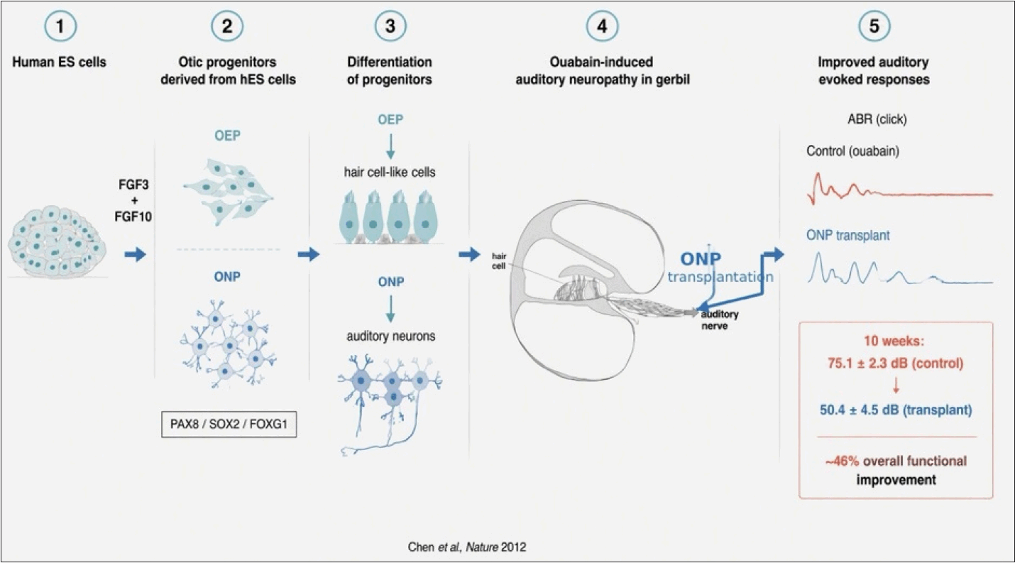
Development and transplantation of human ES cell-derived otic progenitors for auditory neuropathy. Human embryonic stem cells were exposed to FGF3 and FGF10 to induce otic progenitors, including otic epithelial progenitors (OEPs) and otic neural progenitors (ONPs), characterized by the expression of otic markers such as PAX8, SOX2, and FOXG1. OEPs differentiated into hair cell-like cells, whereas ONPs generated auditory neurons. In a gerbil model of ouabain-induced auditory neuropathy, ONPs were transplanted toward the cochlear auditory nerve. The schematic illustrates the cochlear cross-section, the organ of Corti and hair-cell region, and the auditory nerve. Ten weeks after transplantation, auditory brainstem response (ABR) thresholds improved from 75.1±2.3 dB in ouabain-treated control animals to 50.4±4.5 dB in ONP-transplanted animals, corresponding to an approximately 46% overall functional improvement. The diagram summarizes the experimental strategy and does not imply complete hair-cell regeneration. The figure was generated with the assistance of ChatGPT (OpenAI, https://chatgpt.com) using OpenAI’s image generation model on June 15, 2026.
The functional outcome is particularly important. Histological evidence of transplanted-cell survival or increased neuronal density is encouraging but does not necessarily establish restoration of auditory function. The improvement in auditory-evoked responses provided evidence that transplantation could influence the functional auditory pathway. Nevertheless, functional recovery should not be interpreted as proof of complete reconstruction of the native SGN population. The extent to which donor-derived neurons formed ma-ture Type I SGN connections and contributed directly to the auditory response remains an important question.
Targeting the Injured Spiral Ganglion
Anatomical targeting is one of the major obstacles to cochlear cell transplantation [10,11]. SGNs are located within Rosenthal’s canal, whereas most minimally invasive transplantation routes introduce cells into the scala tympani or another cochlear compartment. Zhang, et al. [12] demonstrated that the injured cochlear microenvironment can influence the migration of transplanted neural stem cells. Following SGN degeneration, stromal cell-derived factor-1 (SDF-1/CXCL12) was upregulated in the injured cochlea. Neural stem cells expressing CXCR4 migrated toward regions of increased SDF-1 signaling. This finding provides an important mechanistic basis for targeted cell delivery. Rather than relying solely on the number of transplanted cells, future transplantation strategies may enhance the efficiency of treatment by improving the ability of cells to recognize and migrate toward the injured SGN compartment.
A complementary approach was reported by Zhang, et al. [13] using the cochlear lateral wall as an alternative delivery route. Neural stem cells derived from the olfactory bulb were transplanted through the cochlear lateral wall in a model of SGN degeneration. A proportion of transplanted cells migrated into Rosenthal’s canal and differentiated into neurons. The study is particularly relevant because it addressed one of the major practical limitations of intracochlear transplantation: the difficulty of getting cells to the location where SGNs reside. The lateral-wall approach provided a relatively direct route to Rosenthal’s canal and demonstrated that the cochlear architecture may permit migration of cells from the lateral wall toward the spiral ganglion. Together, these studies demonstrate that successful SGN regeneration depends on both cell identity and anatomical targeting.
Human Dermal Fibroblast-Derived Induced Neural Stem Cells
Chang, et al. [9] recently reported a further development in this field using induced neural stem cells generated directly from human dermal fibroblasts (Fig. 2). The study generated human dermal fibroblast-derived induced neural stem cells (HDF-iNSCs) using transcription-factor-mediated direct conversion and characterized the cells as neural stem/pro-genitor cells. The cells expressed neural stem-cell-associated markers including Nestin, Sox2, and Pax6. This approach differs conceptually from ESC- or iPSC-based strategies. Instead of first generating pluripotent cells and subsequently directing them toward a neural or otic lineage, somatic fibroblasts are directly converted into neural stem-like cells. This may simplify the manufacturing pathway and potentially reduce some concerns associated with pluripotent intermediates. Following scala tympani transplantation in an auditory-neuropathy model, transplanted HDF-iNSCs survived within the cochlea and were detected in the SGN region, including Rosenthal’s canal. The study reported increased neural structures, including neurons and nerve fibers, and transplanted cells demonstrated neuronal characteristics through expression of neuronal markers including TUJ1, MAP2, and NeuN. These findings are particularly relevant to the concept of SGN neuronal density. The study provides evidence that a human somatic-cell-derived neural progenitor population can increase the neural cellular component of an injured cochlea. Importantly, however, the study went beyond histological evaluation. Central auditory synaptic markers were increased following transplantation, and electrically evoked auditory brainstem responses were significantly enhanced. This finding is highly relevant to cochlear implantation because cochlear implants electrically stimulate auditory neurons rather than restoring the damaged hair-cell sensory apparatus. An increased population of electrically responsive auditory neurons could therefore provide an improved neural substrate for cochlear implant stimulation. At the same time, the findings demonstrate that neural regeneration is incomplete. Peripheral synaptic connections were not fully restored, and conventional acoustic auditory thresholds did not show significant improvement. Therefore, the HDF-iNSC study should not be interpreted as complete restoration of the peripheral auditory pathway. Rather, it provides evidence for a more limited but potentially important biological effect:
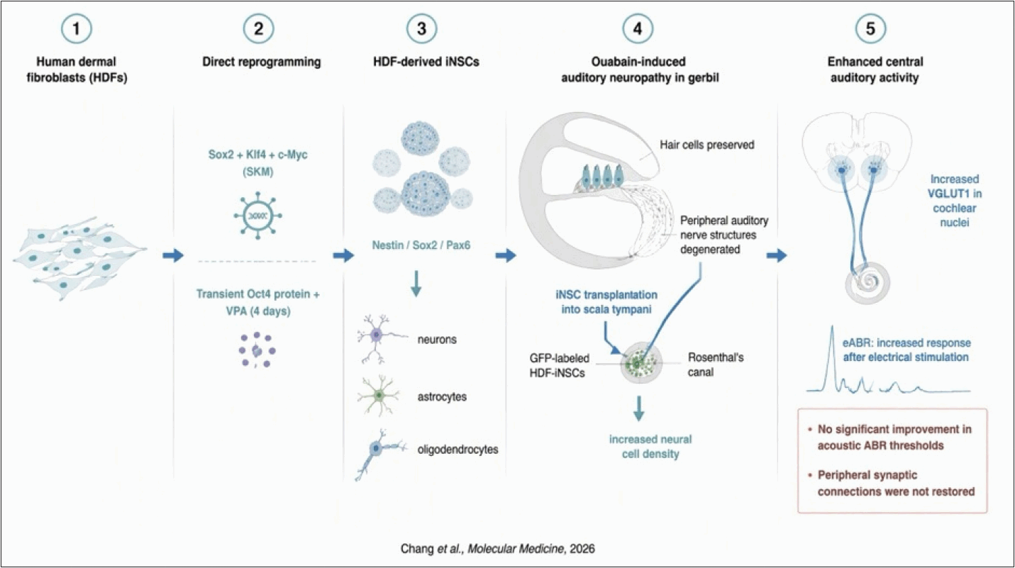
Graphical summary of direct conversion of human dermal fibroblasts into iNSCs and their transplantation in an auditory neuropathy model. Human dermal fibroblasts (HDFs) were directly reprogrammed into induced neural stem cells (iNSCs) using Sox2, Klf4, and c-Myc (SKM), together with transient Oct4 protein and valproic acid (VPA) treatment. The HDF-derived iNSCs expressed Nestin, Sox2, and Pax6 and showed multilineage differentiation potential toward neurons, astrocytes, and oligodendrocytes. In an ouabain-induced auditory neuropathy model in Mongolian gerbils, hair cells were preserved, whereas peripheral auditory nerve structures were degenerated. GFP-labeled HDF-iNSCs were transplanted into the scala tympani through the round window, resulting in increased neural cell density within the cochlea, including the region of Rosenthal’s canal. Transplantation was associated with increased VGLUT1 expression in the cochlear nuclei and enhanced electrically evoked auditory brainstem responses (eABRs). However, acoustic ABR thresholds did not show a significant improvement, and peripheral synaptic connections were not restored. Thus, HDF-iNSC transplantation enhanced neural structure and central auditory activity without demonstrating complete peripheral auditory nerve or synaptic regeneration. The figure was generated with the assistance of ChatGPT (OpenAI, https://chatgpt.com) using OpenAI’s image generation model on June 15, 2026.
Increased cochlear neural structure + enhanced central auditory connectivity + improved electrically evoked neural response.
This combination makes the study particularly relevant to the future development of regenerative strategies designed to complement cochlear implantation.
SGN Neuronal Density as an Intermediate Therapeutic Endpoint
The studies reviewed above suggest that SGN neuronal density may be an important intermediate endpoint in auditory neural regeneration. Parker, et al. [5] demonstrated an increase in Type I SGN-associated cells after neural stem-cell transplantation. Cho, et al. [7] reported increased SGN numbers following transplantation of neural-differentiated human MSCs. Chen, et al. [8] demonstrated that human otic progenitors could differentiate toward auditory neuronal phenotypes and improve auditory-evoked responses. Most recently, Chang, et al. [9] demonstrated increased neural structures and central auditory connectivity following HDF-iNSC transplantation.
However, neuronal density alone should not be considered the final therapeutic endpoint. A transplanted cell located within Rosenthal’s canal does not necessarily constitute a mature Type I SGN. Likewise, expression of general neuronal markers such as TUJ1, MAP2, or NeuN does not establish that the cell has acquired the specialized molecular, electrophysiological, and connectivity characteristics of a mature auditory neuron.
A successful regenerative strategy should ideally accomplish several sequential steps:
1) Survival of transplanted cells.
2) Migration to the SGN compartment.
3) Differentiation toward an appropriate auditory neuronal phenotype.
4) Increase or preservation of the viable SGN population.
5) Extension of peripheral and central neurites.
6) Establishment of functional synaptic connections.
7) Electrical responsiveness compatible with cochlear implant stimulation.
Accordingly, SGN neuronal density should be regarded as an intermediate biological endpoint rather than the ultimate definition of auditory regeneration.
Functional Relevance to Cochlear Implantation
The clinical rationale for SGN regeneration is closely linked to cochlear implantation. Cochlear implants bypass damaged hair cells and directly stimulate the auditory nerve. Consequently, the quantity and quality of surviving SGNs may influence the neural substrate available for electrical stimulation. If cell transplantation can increase the number of viable auditory neurons within Rosenthal’s canal, several potential benefits may follow. These could include increased neuronal recruitment, improved electrode-neural interaction, and potentially more efficient transmission of electrically evoked signals to the central auditory system. The HDF-iNSC study provides particularly relevant evidence because it evaluated electrically evoked auditory responses. The increase in eABR responses after transplantation suggests that the biological changes induced by cell transplantation were associated with an electrically responsive auditory pathway. However, it remains unclear how much of this effect was attributable to newly differentiated donor-derived neurons versus preservation of endogenous neurons or enhancement of central auditory connectivity. This distinction is important for future clinical translation. The objective may not necessarily need to be complete anatomical reconstruction of the native auditory pathway. A more realistic therapeutic goal may be to generate or preserve a sufficiently dense population of mature, electrically excitable neurons capable of effective interaction with a cochlear implant.
Remaining Challenges
Several major challenges remain before SGN-directed cell transplantation can be translated into clinical therapy. First, the identity of regenerated neurons must be established more rigorously. General neuronal markers are insufficient to prove authentic SGN differentiation. Future studies should combine lineage tracing, transcriptomic profiling, electrophysiology, and projection mapping to demonstrate acquisition of a genuine auditory neuronal phenotype. Second, the relative contributions of donor-derived and endogenous neurons remain unclear. Increased SGN numbers may reflect direct donor-cell replacement, trophic preservation, reduced secondary degeneration, or combinations of these mechanisms. Third, delivery remains a major limitation. Rosenthal’s canal is anatomically difficult to access, and conventional scala tympani delivery results in substantial loss of transplanted cells before they reach the SGN compartment. The SDF-1/CXCR4 findings and the cochlear lateral-wall approach suggest potential solutions, but their safety and reproducibility require further study [12,13]. Fourth, neuronal integration must be demonstrated. The ultimate objective is not simply to increase cell number but to establish a functional neural network. Peripheral neurite extension, synaptic formation, central auditory projections, and electrical responsiveness must all be considered. Fifth, long-term safety remains essential. Directly converted iNSCs may have advantages compared with pluripotent stem cells, but prolonged surveillance for abnormal proliferation, phenotypic instability, inappropriate differentiation, and immune responses will be necessary. Finally, the relationship between regenerated neurons and cochlear implant electrodes needs to be investigated. The optimal spatial distribution of regenerated neurons may be different from the distribution required for conventional biological auditory regeneration because cochlear implants directly activate neurons through electrical fields (Fig. 3).
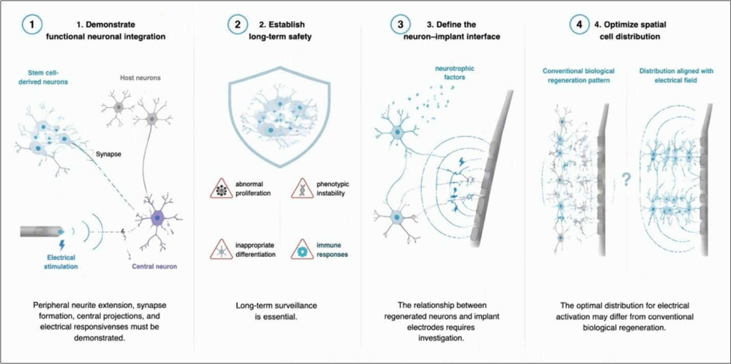
Remaining challenges in stem cell-derived neuronal repair for auditory rehabilitation. Schematic overview of the major challenges that must be addressed to advance stem cell-derived neuronal therapies. 1) Functional neuronal integration: Stem cell-derived neurons must demonstrate peripheral neurite extension, synapse formation with host neurons, central projections, and responsiveness to electrical stimulation. 2) Long-term safety: extended surveillance is required to evaluate abnormal proliferation, phenotypic instability, inappropriate differentiation, and immune responses after transplantation. 3) Neuron–implant interface: the functional relationship between regenerated neurons and implant electrodes, including their response to electrical stimulation and possible interactions with neurotrophic factors, remains to be defined. 4) Spatial cell distribution: the optimal spatial arrangement of regenerated neurons for direct electrical activation may differ from the distribution required for conventional biological regeneration. Together, these challenges highlight the need for further functional, safety, and circuit-level validation of stem cell-derived neuronal therapies. The figure was generated with the assistance of ChatGPT (OpenAI, https://chatgpt.com) using OpenAI’s image generation model on June 15, 2026.
Future Perspective: From Cell Transplantation to an Integrated Regenerative Cochlear Implant
The progression of the field can be viewed as a series of increasingly integrated therapeutic strategies. Early transplantation studies emphasized survival and trophic support. Subsequent studies demonstrated migration toward Rosenthal’s canal and modulation of SGN number. Human MSCs and human ESC-derived otic progenitors [14] introduced humancell and lineage-directed approaches, while chemotactic and alternative delivery strategies addressed the anatomical problem of targeting the SGN compartment. The recent HDF-iNSC study further integrated neuronal regeneration with central auditory connectivity and electrically evoked neural responses. The next stage is likely to combine these approaches rather than rely on cell transplantation alone. An ideal regenerative platform may incorporate lineage-directed or directly converted human neural progenitors, targeted intracochlear delivery, neurotrophic support, modulation of the cochlear microenvironment, and electrical stimulation through a cochlear implant [2,15]. Such an approach could transform the conceptual role of the cochlear implant. Instead of functioning solely as an electrical prosthesis that stimulates a pre-existing population of surviving neurons, the implant could become part of an integrated regenerative system in which biological therapy increases and optimizes the neural substrate available for electrical stimulation.
This concept is particularly attractive because complete reconstruction of the native organ of Corti may not be necessary for clinically meaningful benefit. If a sufficient population of mature, electrically excitable neurons can be generated or preserved within an anatomically appropriate region, the cochlear implant may be able to utilize this regenerated neural substrate. The HDF-iNSC findings provide an important proof-of-concept for this direction. The combination of increased neural structures, enhanced central auditory connectivity, and increased eABR responses suggests that cellular regeneration and electrical stimulation may eventually be developed as complementary rather than independent therapies.
Conclusion
Early neural stem-cell studies established the feasibility of cellular transplantation and suggested trophic effects [4]. Subsequent work demonstrated migration toward Rosenthal’s canal and modulation of Type I SGNs [5]. Human MSC transplantation provided evidence for increased SGN numbers [7], while Chen, et al. [8] demonstrated that human ESC-derived otic progenitors could generate auditory neuronal phenotypes and improve auditory-evoked responses. The development of targeted migration strategies further demonstrated that the cochlear microenvironment and transplantation route are critical determinants of therapeutic success [12,13].
The convergence of human stem-cell technology, targeted intracochlear delivery, neural differentiation, and cochlear implant technology may therefore lead to an integrated regenerative cochlear implant platform, in which biological neural regeneration and electrical auditory prostheses work together to overcome the limitations imposed by progressive SGN degeneration.
Notes
Acknowledgments
This research was supported by the Basic Science Research Program through the National Research Foundation of Korea (NRF) (RS-2020-NR049585; RS-2025–25414419; RS-2026-25619632), and the Bio&Medical Technology Development Program of the National Research Foundation (NRF) funded by the Korean government (RS-2023–00220408).