A Case of Superior Orbital Subperiosteal Abscess Secondary to a Paranasal Sinus Mucocele: Drainage Using a Combined Endoscopic and External Approach
부비동 점액낭종의 합병증으로 발생한 상안와 골막하 농양 1예: 내시경 및 외부접근법을 통한 배농
Article information

Abstract
Superior orbital subperiosteal abscess is a rare but vision-threatening complication of sinusitis or paranasal sinus mucoceles. Due to anatomical constraints, endoscopic drainage alone may be insufficient. We report a case of superior orbital subperiosteal abscess secondary to a mucocele, which was successfully managed with a combined endoscopic and external approach. A 58-year-old male with a history of endoscopic sinus surgery presented with the left orbital swelling and restricted extraocular movement. Imaging revealed a superior orbital subperiosteal abscess associated with an ethmoid sinus mucocele. The patient underwent simultaneous endoscopic sinus surgery for marsupialization of the mucocele and an external approach for drainage of the superior orbital abscess. Postoperatively, the patient remained free of recurrence and had no sequelae during follow-up. In cases of superior orbital subperiosteal abscesses, especially those with mucoceles, distorted anatomy, or previous sinus surgery, a combined endoscopic and external surgical approach may facilitate complete drainage and favorable long-term outcomes.
Introduction
Orbital subperiosteal abscess is a severe orbital complication of acute rhinosinusitis. It may cause visual impairment, restricted extraocular movements, and proptosis and, in severe cases, may lead to intracranial complications, making it a medical emergency [1]. Most orbital subperiosteal abscesses are associated with ethmoid sinusitis and develop along the medial orbital wall. In rare cases, however, they develop along the superior orbital wall in association with frontal or frontoethmoidal sinus disease. Because the superior orbital compartment is anatomically confined, lesions in this location can cause severe pain, inferior displacement of the globe, and optic nerve compression early in the disease course. Superior orbital subperiosteal abscesses may therefore require more complex and aggressive surgical management than medial orbital subperiosteal abscesses [2,3].
Paranasal sinus mucoceles result from mucus retention caused by chronic obstruction of the drainage pathway and gradually expand, eroding the surrounding bone. Mucoceles in the frontal and frontoethmoidal sinuses carry a particularly high risk of extension into the orbit or skull base [4,5]. Although an infected mucocele rarely extends into the orbit and causes a subperiosteal abscess, prompt drainage is essential when this occurs because of the risk of a rapid increase in intraorbital pressure and optic nerve injury [4,6]. When a superior orbital subperiosteal abscess is associated with a mucocele, selecting an appropriate surgical approach may be difficult because of the lesion’s location and the distorted anatomy.
Management of an orbital subperiosteal abscess includes broad-spectrum antibiotics and is tailored according to the abscess size and location, the presence of visual dysfunction, and comorbidities [7]. Recent advances in endoscopic sinus surgery have enabled medial orbital subperiosteal abscesses to be drained using a relatively less invasive approach. However, an endoscopic approach alone may not provide adequate visualization or a sufficient drainage pathway for a superior orbital abscess. An external or combined approach may be necessary, particularly when a mucocele is present or previous surgery has caused substantial anatomical distortion [2,5,6].
We encountered a patient with a superior orbital subperiosteal abscess associated with a mucocele that developed after previous endoscopic sinus surgery. The patient was successfully treated using simultaneous endoscopic and external approaches. We report this case with a review of the literature and discuss the utility of a combined approach for superior orbital subperiosteal abscesses.
Case
A 58-year-old male with diabetes mellitus, hypertension, and asthma had undergone bilateral endoscopic sinus surgery for chronic rhinosinusitis with nasal polyps at another hospital 10 years earlier. After experiencing recurrent periods of improvement and worsening, he presented to our outpatient clinic with left periorbital swelling that had developed that morning (Fig. 1A). His diabetes mellitus was well controlled, with an HbA1c level of 5.6% and a normal blood glucose level. His asthma was asymptomatic and well controlled with a low-dose inhaled corticosteroid. Nasal endoscopy showed postoperative changes from the previous bilateral endoscopic sinus surgery and recurrent nasal polyps. Computed tomography (CT) of the paranasal sinuses (Fig. 2) and magnetic resonance imaging (MRI) (Fig. 3) showed an ethmoid sinus mucocele with erosion and involvement of the superior orbital wall and skull base, and a left superior orbital subperiosteal abscess was suspected. Visual acuity was 1.0 in the right eye and had decreased to 0.7 in the left eye, and the patient reported diplopia with left hypotropia. Ophthalmologic evaluation showed restricted extraocular movements in all directions in the left eye, along with chemosis and conjunctival injection. Intraocular pressure was normal bilaterally, and no relative afferent pupillary defect was present. Based on these findings, he was diagnosed with a superior orbital subperiosteal abscess associated with a mucocele. In consultation with the ophthalmology department, we planned abscess drainage under general anesthesia using endoscopic sinus surgery combined with an external approach.
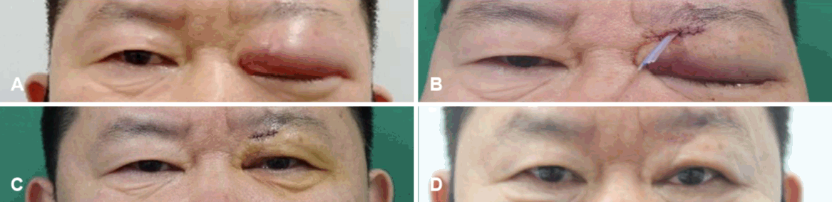
Clinical photographs of a 58-year-old male. A: Preoperative appearance. B: Postoperative state (postoperative day [POD] #0). C: Postoperative state after drain removal (POD #2). D: Postoperative state after stitch out (POD #11).
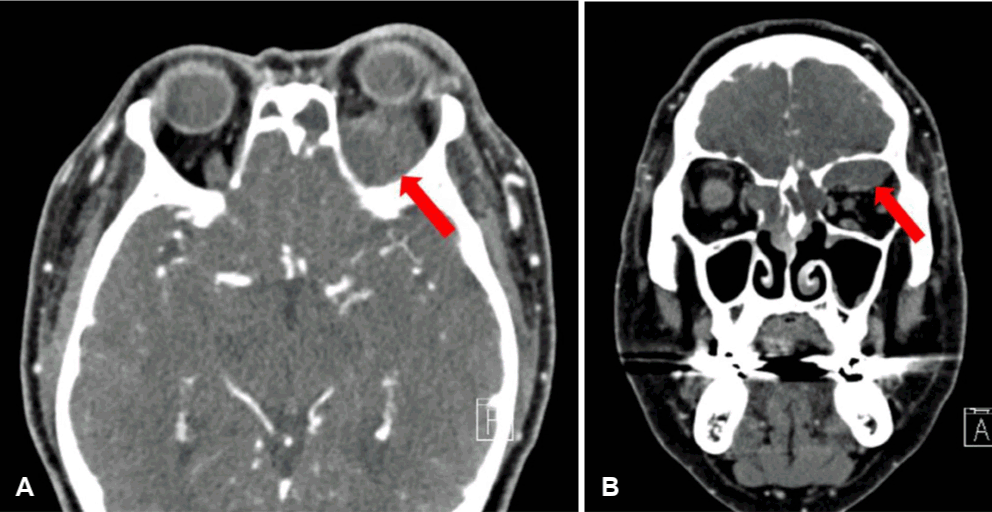
Preoperative contrast-enhanced facial computed tomography showing left superior subperiosteal abscess (red arrow) with ethmoid mucocele and periorbital cellulitis. A: Axial view. B: Coronal view.
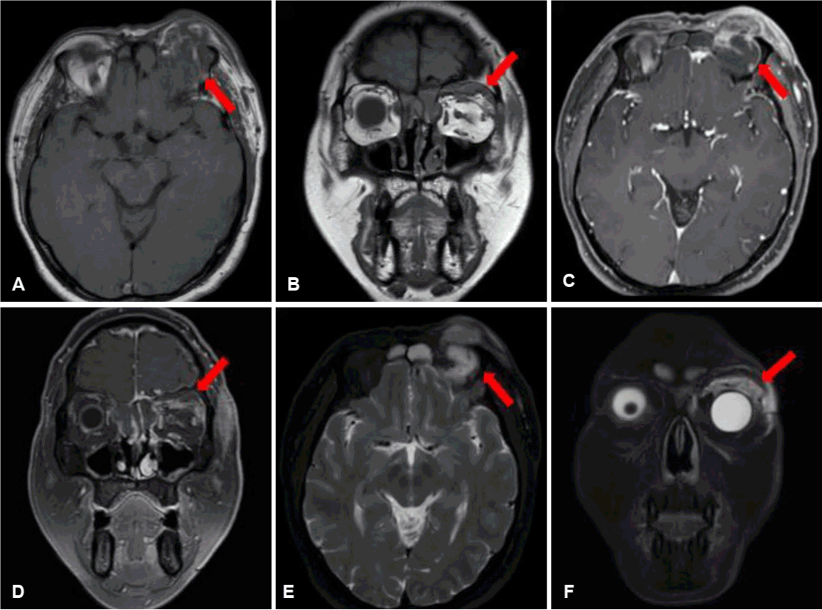
Preoperative magnetic resonance imaging showing left superior subperiosteal abscess (red arrow) with ethmoid mucocele (A) Axial T1, (B) Coronal T1, (C) Axial T1 Enhance, (D) Coronal T1 Enhance, (E) Axial T2, and (F) Coronal T2.
Intraoperatively, the maxillary sinus ostia were patent bilaterally. Multiple nasal polyps around the nasal septal mucosa were resected. The frontal intersinus septum had been eroded by the mucocele, partially connecting the two frontal sinuses. The mucocele contained mucopurulent material on the left and relatively serous material on the right. After the posterior aspect of the left superior orbit was approached endoscopically, the opening into the lesion was enlarged, allowing pus and bloody discharges to drain (Fig. 4A). However, the superior orbital space was too narrow and deep to maintain an adequate drainage pathway using an endoscopic approach alone. The ophthalmology team therefore added an external approach to drain the superior orbital abscess more definitively, and a Silastic drain was placed (Fig. 1B).
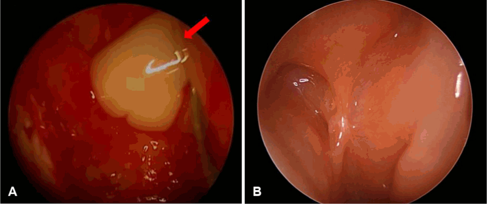
Left nasal endoscopic findings. A: Intraoperative endoscopic view of pus drainage (red arrow) from left subperiosteal orbital abscess. B: Postoperative endoscopic view at 5 years showing a widely patent mucocele marsupialization site without evidence of recurrence.
Intravenous antibiotics were administered postoperatively, and culture yielded Klebsiella aerogenes. The left periorbital swelling and ophthalmologic symptoms gradually improved, and the Silastic drain was removed on postoperative day 2 (Fig. 1C). Paranasal sinus CT performed on postoperative day 4 showed that the left superior orbital subperiosteal abscess had been adequately drained, with a marked reduction in lesion size (Fig. 5A and B). The patient was discharged without complications. At the ophthalmologic examination on postoperative day 11, visual acuity had recovered to 1.0 in the left eye. No restriction of extraocular movements was observed in any direction in the left eye, and the diplopia had completely resolved. The patient has been followed as an outpatient for 5 years since surgery without recurrence or ophthalmologic sequelae (Figs. 1D, 4B, and 5C).
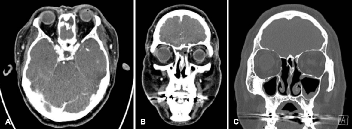
Postoperative contrast-enhanced facial computed tomography. A: Postoperative day (POD) #4, axial view. B: POD #4, coronal view. C: POD #1 year. The images show marked resolution of the superior orbital subperiosteal abscess and ethmoid mucocele after drainage.
This case report was granted an exemption from review by the Institutional Review Board of Hallym University Sacred Heart Hospital (HALLYM 2026-04-048).
Discussion
Superior orbital subperiosteal abscess is a rare orbital complication associated with sinusitis or a mucocele and may cause visual impairment, proptosis, and restricted extraocular movements. Diagnosis and treatment decisions are particularly difficult because superior orbital lesions are deep and access is restricted by their anatomical location [2,3]. In adults, superior orbital subperiosteal abscess secondary to a mucocele is even rarer. Early diagnosis and aggressive treatment are important because this condition can cause permanent visual impairment [8,9].
Paranasal sinus mucoceles are classified as primary or secondary according to their cause. Primary mucoceles develop without a specific predisposing factor, whereas secondary mucoceles may be associated with stenosis or obstruction of the drainage pathway following trauma, chronic inflammation, or previous sinus surgery [3]. In this case, a mucocele with orbital complications developed despite well control of the patient’s underlying diabetes mellitus and asthma. Given his history of endoscopic sinus surgery 10 years earlier, recurrent symptoms thereafter, and imaging findings of expansile bony changes with involvement of the superior orbital wall and skull base, the lesion was considered a secondary mucocele related to the previous surgery rather than to poor control of his comorbidities. Compared with orbital abscesses arising from rhinosinusitis without an associated mucocele, lesions associated with secondary mucoceles may involve erosion of the surrounding bone and anatomical distortion. Accurate preoperative assessment of their extent using CT and MRI is therefore particularly important [4,6-8].
Surgical treatment of an orbital subperiosteal abscess varies according to the lesion’s location and extent, the presence of visual dysfunction, and the underlying condition. Abscesses along the medial orbital wall can generally be adequately drained using an endoscopic approach alone. However, when an abscess is located along the superior or lateral orbital wall, limited endoscopic visualization and instrument maneuverability may preclude complete drainage [2,9]. Moreover, in cases such as this one, in which a mucocele is present, the frontoethmoidal anatomy is markedly distorted by previous surgery, and the lesion extends to the posterior aspect of the superior orbit, it may be difficult to maintain a stable and adequate drainage pathway using an endoscopic approach alone. A combined approach that includes an external procedure may therefore be a more effective treatment strategy [5,6].
Previous reports of superior orbital subperiosteal abscesses or lesions associated with frontal sinus mucoceles have primarily used an external approach. Other cases required a secondary external approach because improvement was insufficient after initial endoscopic drainage [4,5]. Recent reports (Table 1) indicate that simultaneous endoscopic and external approaches to superior lesions provide a wider operative field and reliable drainage, with low recurrence rates [4,5,10-13]. Most of the available evidence, however, consists of case reports or small case series, making it difficult to conclude that the combined approach is directly superior. Nevertheless, when factors such as a superior orbital lesion, an associated mucocele, or anatomical distortion from previous surgery are present, it may be reasonable to consider a combined approach from the outset.
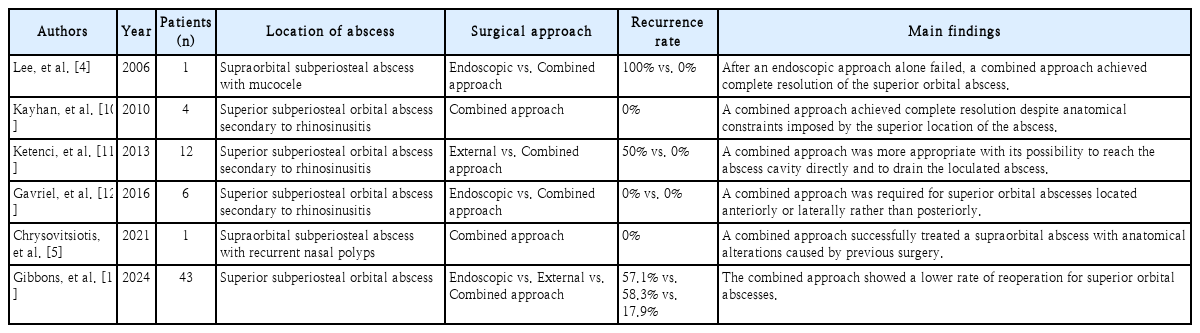
Comparison of previous studies on surgical approaches for superior orbital subperiosteal abscesses
In this case, we marsupialized the sinonasal mucocele endoscopically and simultaneously added an external approach to drain the superior orbital abscess directly and adequately. A drain was placed to maintain the drainage pathway. This strategy was intended to reduce the risks of inadequate drainage and reoperation associated with an endoscopic approach alone. The patient’s ophthalmologic symptoms improved postoperatively, and he had a favorable course without recurrence or ophthalmologic sequelae during long-term follow-up. This case, together with the literature review, suggests that an upfront combined approach may be effective for superior orbital subperiosteal abscesses, particularly when a mucocele or severe anatomical distortion is present.
In conclusion, this case describes successful treatment of a superior orbital subperiosteal abscess caused by a secondary paranasal sinus mucocele through simultaneous endoscopic and external approaches. A combined surgical approach may be effective when anatomical factors such as a superior abscess location, an associated mucocele, or prior surgery are present. This case may support the clinical utility of this strategy.
Notes
Acknowledgments
None
Author Contribution
Conceptualization: Jee Hye Wee. Data curation: Kyung Hoon Park, Geun Sang Lee. Visualization: Kyung Hoon Park. Writing—original draft: Kyung Hoon Park, Geun Sang Lee. Writing—review & editing: Ming Joung Lee, Jee Hye Wee. Supervision: Jee Hye Wee.