Oral Tuberculosis Mimicking Oral Cavity Cancer With Pulmonary Metastasis: A Case Report
폐전이를 동반한 구인두암으로 오인된 구강 결핵 1예
Article information

Abstract
Oral tuberculosis (TB) is an uncommon manifestation, accounting for only a small fraction of extrapulmonary TB cases and often mimicking malignant lesions by its chronic ulcerative or mass-forming appearance. Such presentations frequently lead to misdiagnosis as squamous cell carcinoma or other head and neck malignancies. When pulmonary lesions coexist, distinguishing oral TB from metastatic cancer becomes even more challenging. A 41-year-old male presented with a painful oral mass, accompanied by multiple pulmonary nodules on chest CT, raising strong suspicion of primary oral malignancy with lung metastasis. Initial clinical impression favored a malignant disease due to the mass-like appearance of the oral lesion and radiologic features of disseminated pulmonary nodules. However, incisional biopsy of the oral mass revealed granulomatous inflammation, consistent with TB. Similar cases of oral lesions leading to the diagnosis of systemic or pulmonary TB have been reported, emphasizing the importance of tissue confirmation.
Introduction
Oral tuberculosis is a rare disease that typically occurs as a secondary lesion of pulmonary tuberculosis. It may present in various forms, including chronic ulcers and mass-like lesions, making clinical differentiation from malignancy difficult [1]. When oral lesions are accompanied by multiple pulmonary lesions, the condition can easily be mistaken for metastatic head and neck cancer rather than tuberculosis. Here, we report a case of oral tuberculosis initially misdiagnosed as oral cancer and emphasize the importance of including tuberculosis in the initial differential diagnosis in similar clinical presentations.
Case
A 41-year-old male presented with a 3-month history of an intraoral lesion and dysphagia. The patient, originally from Kazakhstan, had been evaluated at a medical institution abroad following symptom onset, where palatal cancer with pulmonary metastasis was suspected. He was subsequently referred to our hospital for further evaluation. He was in good general condition. He reported odynophagia but had no dyspnea or trismus. Physical examination revealed an oval mass with ulceration, measuring 1.5×0.5 cm, located at the junction of the right hard and soft palate (Fig. 1). A 2-cm, firm, painless lymph node was palpated in the left level II region. His medical history was unremarkable, with no underlying diseases or history of tuberculosis.
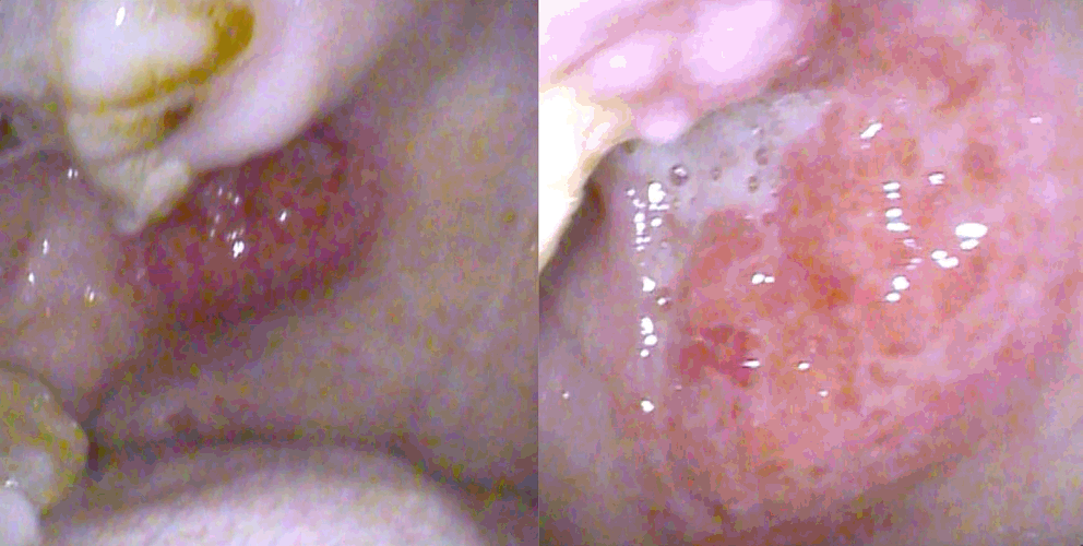
Clinical photograph showing an ulcerative mass lesion at the junction of the right hard and soft palate.
Contrast-enhanced neck CT showed multiple enlarged cervical lymph nodes at the right levels Ib and II (Fig. 2). Neck MRI further demonstrated an ulcerative lesion in the right soft palate, characterized by high signal intensity on T1-weighted images and prominent contrast enhancement; therefore, malignancy could not be excluded (Fig. 3). Positron emission tomography (PET)-CT revealed increased fluorodeoxyglucose (FDG) uptake in the oral and pharyngeal regions, as well as in the cervical lymph nodes. Additionally, multiple small nodules distributed throughout both lungs exhibited increased FDG uptake, raising a strong suspicion of pulmonary metastasis originating from oral cancer (Fig. 4).
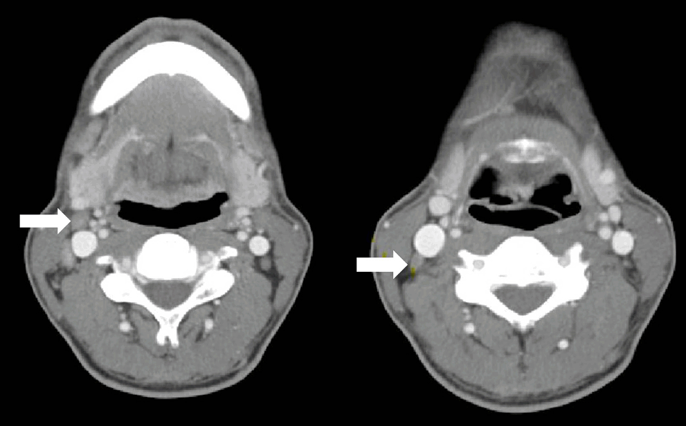
Contrast-enhanced neck CT showing multiple enlarged lymph nodes in the right level Ib and II regions (white arrow: level Ib lymph node and level II lymph node).
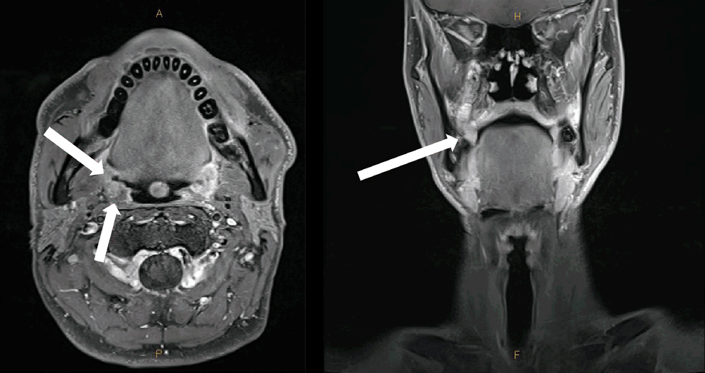
MRI of the neck demonstrates an ulcerative lesion in the right soft palate with prominent contrast enhancement (white arrows).
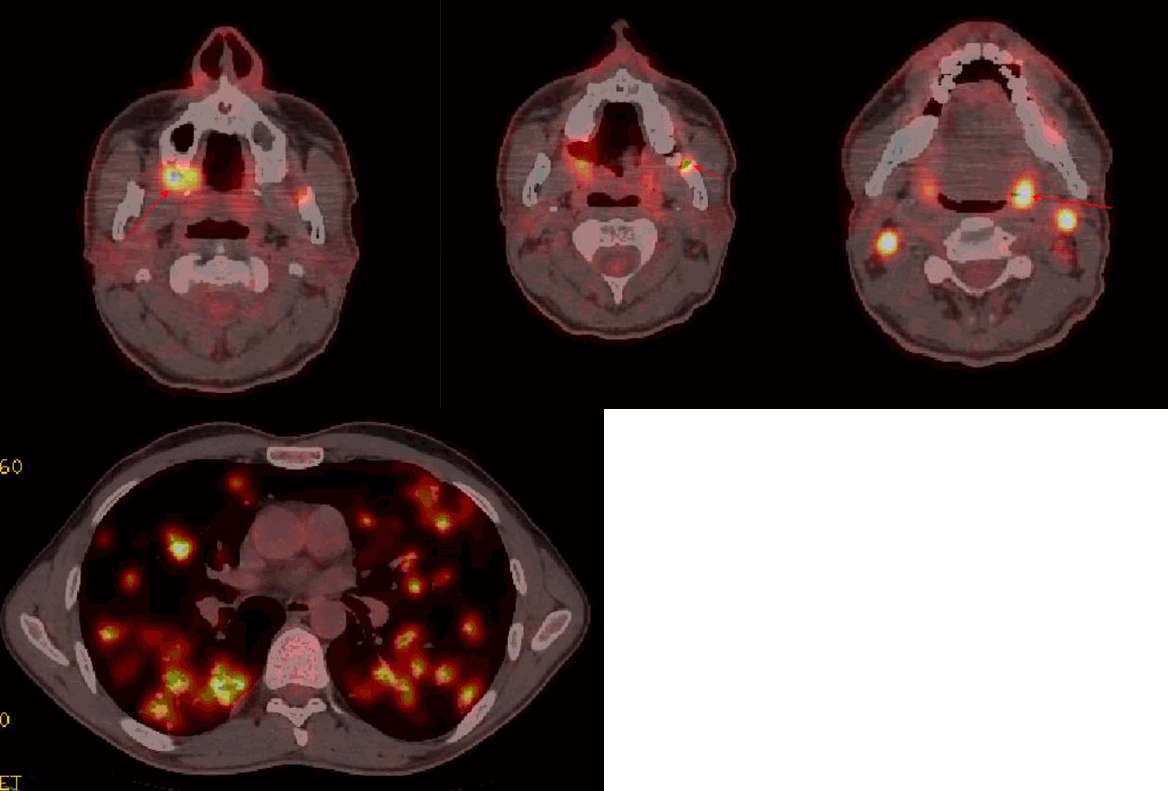
Positron emission tomography–CT revealing increased fluorodeoxyglucose uptake in the oral and pharyngeal regions, as well as multiple pulmonary nodules.
The initial biopsy performed at presentation showed atypical squamous epithelium with dense inflammatory cell infiltration. Due to the severe inflammatory reaction, distinguishing squamous cell carcinoma from regenerative changes was difficult, and oral cancer remained a clinical concern (Fig. 5). Human papillomavirus polymerase chain reaction (PCR) testing was negative. One week later, a repeat biopsy of the lesion was performed to obtain an accurate pathological diagnosis. This repeat biopsy demonstrated an ulcerative lesion with inflammatory exudate, abundant acute and chronic inflammatory cells, and focal regenerative atypical squamous epithelium. Notably, subtle granulomatous inflammation was observed in some tissue fragments, suggesting the possibility of tuberculosis when interpreted alongside the clinical findings (Fig. 6).
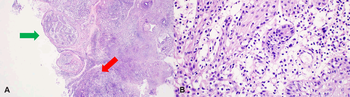
Histopathological findings from the initial biopsy show dense inflammatory cell infiltration (red arrow) with atypical squamous epithelium (green arrow). Hematoxylin & Eosin Staining, A: ×40, B: ×400.
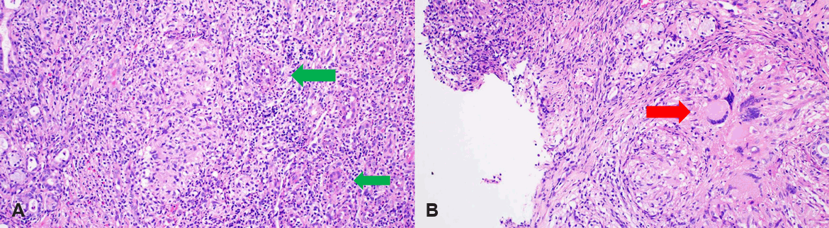
Histopathological findings from the repeat biopsy demonstrating focal granulomatous inflammation without necrosis, suggestive of tuberculosis (green arrows: epithelioid histiocytes, red arrow: Langhans giant cell). Hematoxylin & Eosin Staining, A: ×200, B: ×400.
Based on these histopathological findings, an additional tuberculosis PCR analysis was conducted for differential diagnosis. The test was positive for Mycobacterium tuberculosis DNA and negative for nontuberculous mycobacteria. Acid-fast bacillus (AFB) culture confirmed the growth of Mycobacterium tuberculosis in a liquid medium, although the solid-medium culture and AFB smear were both negative.
An additional chest CT scan revealed multiple small nodular lesions throughout both lungs (Fig. 7), which corresponded to the areas previously suspected of being multiple pulmonary metastases on PET-CT. However, given the positive tuberculosis PCR and culture results, these lesions were reinterpreted as being consistent with pulmonary tuberculosis. The final diagnosis was established as oral tuberculosis with concurrent pulmonary tuberculosis.
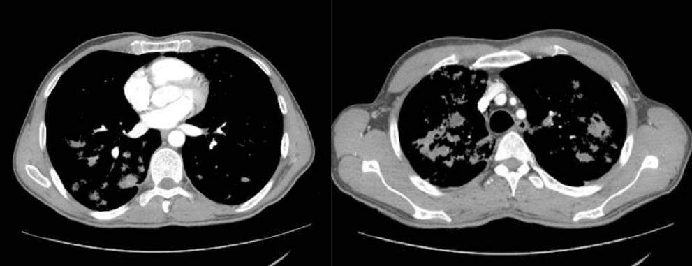
Chest CT showing multiple small nodular lesions throughout both lungs, initially suspected as metastatic lesions and later confirmed as pulmonary tuberculosis.
In this patient, an oral mass, cervical lymph node enlargement, and multiple pulmonary nodules were observed simultaneously, all of which demonstrated increased FDG uptake on PET-CT. Therefore, a careful differential diagnosis was required to determine whether these findings represented malignancy alone or concomitant tuberculosis and malignancy. To improve diagnostic accuracy, the second biopsy included both the central and peripheral portions of the oral mass, and approximately half of the entire lesion was excised. Because no malignant cells were detected in this specimen, the likelihood of occult malignancy in the residual lesion was considered low. Furthermore, although PET-CT showed increased FDG uptake in the cervical lymph nodes, concurrent neck MRI showed that the lymph node enlargement was not severe. Based on their size and morphology, the nodes were interpreted as reactive rather than malignant. Consequently, the possibility of oral cancer with cervical lymph node involvement and distant pulmonary metastasis was largely excluded, supporting a final diagnosis of oral tuberculosis accompanied by pulmonary tuberculosis.
Nine months after the initial diagnosis, a telephone follow-up was conducted after the patient had returned to his home country. He was continuing his anti-tuberculosis medication regimen, his overall health status was good, and the oral lesion had decreased in size without any surgical intervention. This clinical improvement further supported the exclusion of underlying malignancy. He was confirmed to be taking second-line tuberculosis drugs, including bedaquiline, cycloserine, linezolid, clofazimine, and prothionamide. Although drug susceptibility testing for Mycobacterium tuberculosis was not performed at our hospital, it appears it was conducted at a local hospital in Kazakhstan, where multidrug-resistant tuberculosis (MDR-TB) was confirmed, prompting the prescription of these specific medications. The inability to obtain follow-up imaging from the local hospital remains a limitation of this case report.
Additionally, previously reported cases of oral tuberculosis that were initially misdiagnosed as malignancies are summarized in Table 1.
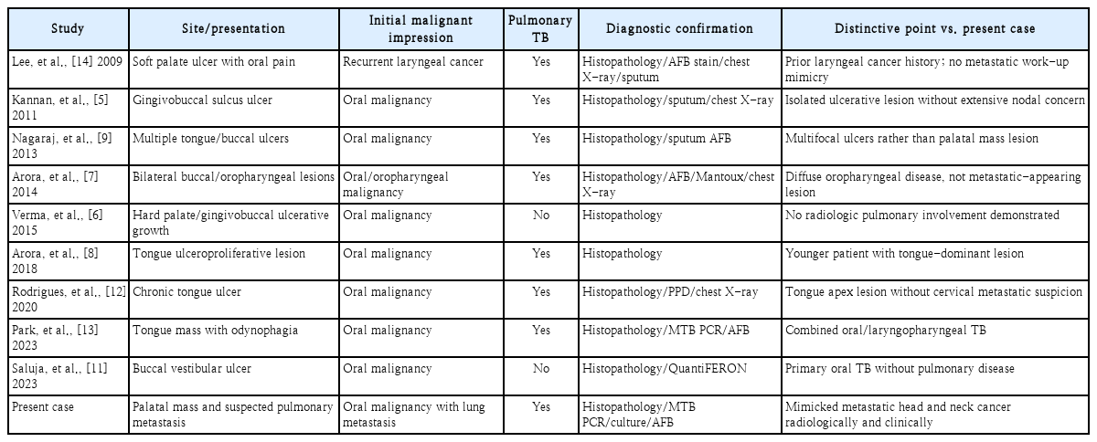
Previously reported cases of oral tuberculosis initially misdiagnosed as malignancy and comparison with the present case
Discussion
Oral tuberculosis is a rare manifestation that accounts for a minimal proportion of all tuberculosis cases and typically occurs as secondary involvement from pulmonary tuberculosis [1-3]. Intraoral lesions can present in various forms, including chronic ulcers, nodules, masses, and hypertrophic lesions. These nonspecific features often complicate the clinical differentiation from malignancy [4-8]. Mass-forming lesions, in particular, are easily mistaken for squamous cell carcinoma, and several cases have been reported where lesions initially suspected to be cancer were ultimately confirmed as tuberculosis only after repeated biopsies or extensive metastatic evaluations [5-8].
In the present case, the patient exhibited both an ulcerative intraoral mass and multiple pulmonary nodules. This clinical presentation is a classic scenario that initially raises suspicion for pulmonary metastasis from head and neck cancer, a common pattern of misdiagnosis documented in the literature [9,10]. While high FDG uptake on PET-CT further heightened the suspicion of malignancy, tuberculosis can yield similar findings due to inflammatory reactions and macrophage activation. Therefore, imaging alone is insufficient for a definitive differential diagnosis [9,10].
The difficulty in distinguishing malignancy from regenerative changes during the initial biopsy, primarily due to intense inflammation, aligns with previous reports [8,11,12]. In some instances, the diagnosis is delayed because distinct granulomas are absent in the initial tissue specimens. In our case, a limited number of subtle granulomatous inflammatory foci were identified only in the second incisional biopsy, and tuberculosis was ultimately confirmed by molecular testing and culture. These findings underscore that tuberculosis cannot be excluded based solely on histological features; rather, clinical, radiological, and molecular diagnostic data must be integrated.
Previous literature has highlighted cases in which an oral lesion was the first indicator of systemic tuberculosis, or in which pulmonary tuberculous lesions were misidentified as malignancy [12-14]. However, cases in which an oral mass and multiple pulmonary nodules are present simultaneously, leading to a strong initial suspicion of metastatic cancer before tuberculosis is confirmed, are relatively rare. The present case illustrates this diagnostic pitfall and emphasizes that infectious diseases, particularly tuberculosis, should be included in the differential diagnosis when oral lesions of unknown etiology are accompanied by pulmonary involvement.
The patient’s epidemiological background is also relevant. Due to time and financial constraints, anti-tuberculosis treatment could not be initiated at our institution, and the patient returned to Kazakhstan for care. The incidence of tuberculosis in Kazakhstan is approximately 70 cases per 100000 population, which is about twice the rate in South Korea (33 cases per 100000). Furthermore, due to healthcare-system challenges in Kazakhstan and neighboring regions, MDR-TB is highly prevalent among newly diagnosed patients, accounting for an estimated 20% to 30% of cases [15]. Consistent with this, our patient was prescribed second-line anti-tuberculosis drugs suitable for MDR-TB at his local hospital, rather than standard first-line therapies. Treatment for MDR-TB typically spans 6 to 9 months, though it may be extended to 12 or 18 months depending on disease severity and clinical response. While the treatment success rate for drug-susceptible tuberculosis is around 90%, the success rate for MDR-TB is notably lower, at approximately 60% to 70%.
In conclusion, oral tuberculosis is a rare infectious disease that can clinically and radiologically mimic malignancy, and when concomitant pulmonary nodules are present, it can easily be misdiagnosed as metastatic cancer. In this case, malignancy was strongly suspected due to the coexistence of an intraoral mass and multiple pulmonary nodules, but tuberculosis was ultimately confirmed through repeated biopsies and molecular diagnostics. Tuberculosis must be included in the differential diagnosis of oral ulcers or masses of unknown origin. Repeated biopsies may be required to secure an accurate diagnosis, and molecular testing for tuberculosis plays an important role.
Notes
AI-assisted Translation
Generative AI was used for English translation. The final version was reviewed and approved by the authors and the Editorial Office, and the authors retain final responsibility for the content.
Bilingual Publication
This article is available in both Korean and English.
Acknowledgments
The authors would like to thank Yeon Hee Kim, MD, for her assistance with pathological consultation and interpretation, as well as for providing and photographing the histopathologic images. We also thank Hyung Kwon Byeon, MD, PhD, for supervision and for his contributions to writing (review and editing).
Author Contribution
Conceptualization: Min Seok Kang. Data curation: Min Seok Kang. Formal analysis: Da Ol Im. Investigation: Da Ol Im. Methodology: Min Seok Kang. Project administration: Min Seok Kang. Resources: Da Ol Im. Software: Da Ol Im. Supervision: Min Seok Kang. Validation: Jae Eun Oh, Gun Ho Kim. Visualization: Da Ol Im, Min Seok Kang. Writing—original draft: Da Ol Im, Gun Ho Kim. Writ ing—review & editing: Jae Eun Oh, Min Seok Kang.