Surgical Reconstruction of Posterior Cricoid Injury Following Airway Management Resulting in Tracheoesophageal Fistula: A Case Report
기도 관리 중 발생한 기관식도누공을 동반한 후윤상연골 손상의 수술적 치료: 증례 보고
Article information

Abstract
Acquired tracheoesophageal fistula (TEF) in adults may arise from iatrogenic injury during airway management; in particular, hypopharyngeal-subglottic fistula (HSF) involving the cricoid is rare. We report a 61-year-old male who underwent repeated intubations and percutaneous tracheostomy for infective endocarditis. During recovery, aspiration led to the diagnosis of a high-level TEF with bilateral vocal fold paralysis (BVFP). Imaging 4 months after tracheostomy suggested posterior cricoid compromise, and surgery revealed fibrosis, loss of cricoid architecture, and a small posterior fistula. Open airway reconstruction using a laryngeal cleft-type multilayer closure with autologous graft achieved durable closure, preserved airway function, and enabled gradual decannulation without recurrence at 1 year. This case highlights that iatrogenic airway injuries can cause delayed posterior cricoid collapse with HSF and BVFP, and that multilayer reconstruction is an effective treatment in complex adult cases.
Introduction
Acquired tracheoesophageal fistula (TEF) in adults is most commonly associated with malignancy, prolonged endotracheal intubation, or tracheostomy-related complications [1]. Specifically, hypopharyngeal-subglottic fistula (HSF) is a high-level TEF involving the cricoid cartilage [2]. HSF involving the cricoid framework is exceedingly rare in adults, and only a limited number of cases have been reported in the literature [2]. Because of its rarity and complex anatomical involvement, diagnosis and surgical management remain challenging.
Cricoid cartilage forms the structural foundation of the posterior larynx and provides critical support for arytenoid stability and vocal fold mobility. Disruption of the cricoid framework may result in mechanical instability, fixation of the cricoarytenoid joints, or secondary neural dysfunction, potentially leading to bilateral vocal fold paralysis (BVFP) [3].
We report a rare case of posterior cricoid injury following airway management presenting with concurrent HSF and BVFP, successfully managed with open surgical reconstruction.
Case
The case was reported based on a prospective registry, using informed consent from patients approved by the Institutional Review Board (No. 2109-043-1253). The written informed consent was obtained from the patient.
A 61-year-old male with a history of treated aortic aneurysm (12 years prior) was hospitalized for infective endocarditis. A summary of the patient’s clinical course is presented in Supplementary Table 1.
Following redo open heart surgery for infective endocarditis, the patient developed leakage at the distal descending aortic anastomosis site approximately 6 weeks postoperatively, resulting in massive hemorrhage requiring extracorporeal membrane oxygenation support. In this setting, percutaneous dilatational tracheostomy (PDT) was performed to facilitate ventilatory management and expedite transfer to the general ward. During the post-intensive care unit recovery period following transfer to the general ward, the patient developed aspiration symptoms and was referred to our department. Initial laryngoscopic examination demonstrated bilateral vocal fold immobility without any definite endoscopic findings suggestive of a tracheoesophageal fistula (Supplementary Fig. 1A). Subsequent videofluoroscopic swallowing study raised suspicion for a tracheoesophageal fistula, which was confirmed by esophagography at the cervical esophageal level (Fig. 1A). Subsequent endoscopic evaluation revealed no obvious posterior wall defect at the level of the tracheostomy tube. However, salivary secretions were observed descending below the vocal folds. Although the tracheostomy site appeared appropriately positioned, these findings raised clinical suspicion of posterior cricoid wall injury or fracture (Fig. 1B and C).
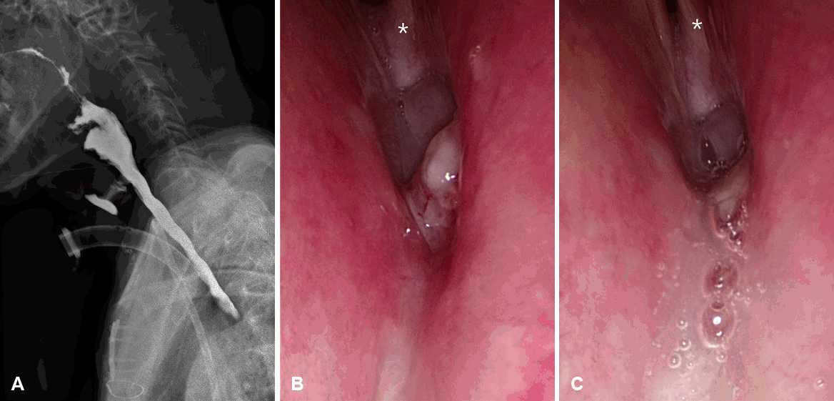
Endoscopic and radiographic findings. A: Esophagography demonstrating a high-level fistula at the C6–7 level. B: Endoscopic view through the percutaneous dilatational tracheostomy stoma, directed cranially toward the vocal folds (asterisk), revealing granulation tissue at the posterior cricoid region. C: Saliva observed passing downward through the defect during swallowing.
Retrospectively, CT obtained 4 months after decannulation following the PDT procedure did not demonstrate any definite structural abnormality of the posterior cricoid region; therefore, the precise timing and mechanism of injury could not be definitively determined. The presumed mechanisms included 1) long-term or traumatic endotracheal intubation, 2) PDT-related injury, or 3) combined iatrogenic structural damage.
Under general anesthesia, a direct airway examination was performed in the operating room. Pooling of saliva was observed immediately below the vocal folds; however, precise assessment of the defect size and location was limited due to restricted visualization caused by cervical rigidity. Because of the patient’s poor general condition following multiple cardiothoracic procedures, definitive repair was deferred. Conservative management with percutaneous endoscopic gastrostomy feeding was continued while monitoring for spontaneous fistula closure with serial esophagography. However, follow-up esophagography demonstrated persistent fistula without interval improvement, and definitive surgical repair was subsequently undertaken.
Accordingly, open airway reconstruction was planned to achieve definitive closure of the fistula and structural stabilization of the posterior cricoid region. Given the location of the defect and the associated posterior cricoid involvement, a surgical approach analogous to type III laryngeal cleft repair was adopted. The operative plan included careful separation of the tracheal and esophageal layers followed by multilayer reconstruction using autologous graft material to reinforce the compromised cricoid framework.
Intraoperative exploration revealed extensive fibrosis replacing the anterior cricoid cartilage; intact or solid cricoid framework could not be clearly identified, and only friable fibrotic tissue was present in the expected location of the anterior cricoid arch (Fig. 2). A small defect measuring approximately 3 mm in diameter was identified at the posterior cricoid wall, consistent with the suspected HSF. The defect was confined to the posterior cricoid region. Careful dissection was performed to separate the tracheal and esophageal layers. Dissection was initiated centrally and extended laterally in an attempt to identify preserved cartilage; however, severe fibrosis limited clear delineation of normal anatomical planes.
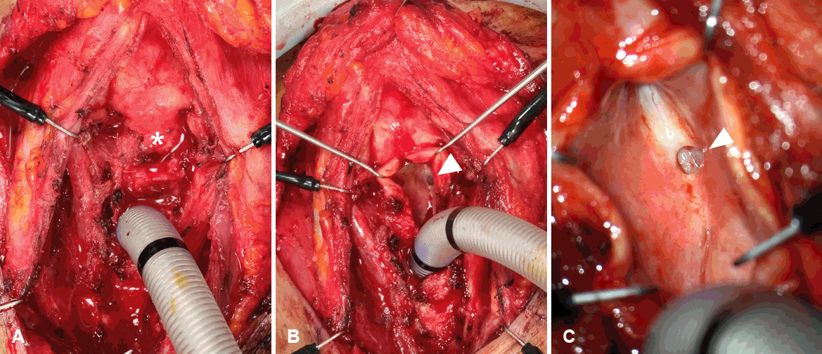
Intraoperative findings. A: At the expected location of the cricoid cartilage (asterisk), only fibrotic and friable tissue was identified in the anterior portion. B: After opening this area, a fistulous defect was visualized at the posterior wall (arrowhead). C: Microscopic magnification of the defect (arrowhead).
After adequate mobilization, approximately a 1×1 cm area was undermined to allow graft placement. Autologous rib cartilage was harvested and inserted with the perichondrium oriented toward the inferior layer to provide structural reinforcement of the posterior cricoid region.
To augment the tracheal side, clavicular periosteum was interposed as an additional reinforcing layer. Multilayer reconstruction was completed, and tissue adhesive was applied to stabilize the graft and closure site.
The patient was maintained on strict nil per os status with gastrostomy tube feeding postoperatively. Oral intake was not recommended until fistula closure had been confirmed by follow-up esophagography. However, the patient independently resumed oral feeding before this evaluation. Followup esophagography revealed no evidence of fistula recurrence (Fig. 3). Following confirmation of fistula closure and the absence of significant aspiration, microlaryngoscopic laser posterior cordotomy was subsequently performed to facilitate decannulation (Supplementary Fig. 1B).
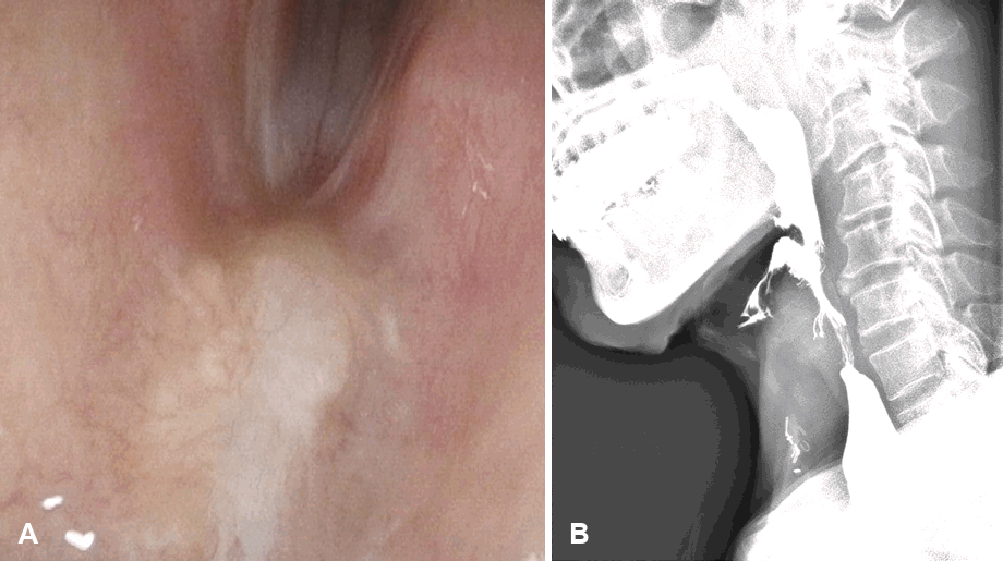
Postoperative findings at 6 months. A: Endoscopic view through the stoma, directed cranially toward the vocal folds, showing the reconstructed surgical site. B: Esophagography demonstrated no evidence of fistula.
After improvement of the glottic airway, gradual T-cannula weaning was carefully initiated. For safety, the tracheostoma was not fully closed, and a small residual stoma was preserved to permit emergency airway access. Following successful T-cannula weaning, the tracheostoma was closed without respiratory compromise. The patient remains under ongoing surveillance.
Discussion
Iatrogenic TEF in adults most commonly results from prolonged endotracheal intubation or tracheostomy-related complications, with pressure necrosis of the posterior tracheal wall representing the predominant pathophysiologic mechanism [4]. In addition, although PDT is generally considered safe, posterior tracheal or cricoid injury remains a recognized but uncommon complication [5,6].
In the present case, both multiple episodes of endotracheal intubation and subsequent PDT were performed, making it difficult to attribute the injury to a single causative event. Either procedure may independently produce posterior wall penetration, cartilage destabilization, or ischemic injury through sustained pressure on the posterior laryngotracheal wall. Notably, even when tracheostomy positioning appears technically appropriate, subclinical cartilage injury may evolve into progressive structural collapse over time, as evidenced by the delayed radiologic findings observed in this case.
What distinguishes this case from previously reported iatrogenic HSF is the extent of posterior cricoid involvement. While posterior tracheal wall injury is a recognized complication of airway instrumentation, complete structural failure of the cricoid framework is exceedingly rare and has been only rarely described in adult literature [7,8]. To our knowledge, this is one of the first reports describing HSF associated with progressive cricoid structural collapse following repeated airway instrumentation in an adult.
In our patient, intraoperative findings revealed extensive replacement of the anterior cricoid cartilage by friable fibrotic tissue, with no identifiable intact cartilaginous framework. This finding suggests that repeated airway instrumentation may have resulted in cumulative mechanical and ischemic injury, followed by progressive fibrotic remodeling. CT obtained after PDT demonstrated pericicoid air density, indicating that structural compromise had already occurred by that time; however, the full extent of the structural deterioration became apparent only during surgical exploration.
The concurrence of HSF and BVFP in this patient warrants particular attention. The cricoid cartilage serves as the structural foundation of the posterior larynx, providing essential support for arytenoid stability and posterior glottic integrity. Disruption of this framework may result in mechanical fixation of the cricoarytenoid joints, posterior glottic scarring, or secondary dysfunction of the recurrent laryngeal nerves through perineural fibrosis or traction injury. Despite this anatomical relationship, cricoid structural injury as a cause of BVFP in adults has rarely been described in the context of airway instrumentation [9]. In this case, BVFP was confirmed; although laryngeal electromyography demonstrated preserved neuromuscular function, the relative contributions of mechanical and neural mechanisms could not be fully delineated.
The simultaneous occurrence of HSF and BVFP therefore suggests a global failure of the posterior laryngeal framework, and clinicians should maintain heightened suspicion for vocal fold dysfunction when posterior cricoid injury is identified or suspected.
From a surgical standpoint, this case illustrates both the applicability and the limitations of established laryngeal reconstruction principles in the setting of acquired adult airway injury. For HSF, endoscopic repair using an occluder device has been reported in the literature [2]; however, this technique was not available at our institution. In addition, endoscopic visualization of the defect was limited due to cervical and laryngeal rigidity, making adequate exposure difficult for endoscopic procedure.
Given the posterior cricoid location of the fistula and the associated structural compromise, a surgical approach analogous to type III laryngeal cleft repair was adopted, with multilayer reconstruction using autologous rib cartilage and clavicular periosteum [10]. While laryngeal cleft repair principles have been well described in pediatric literature, their application to adult acquired HSF with circumferential fibrosis presents considerable technical challenges. Severe scarring and loss of normal anatomical planes significantly complicated dissection and graft placement in the present case. The use of autologous structural grafts with interposed vascularized tissue layers represents a sound reconstructive strategy under these circumstances, and fistula was closed successfully. These findings suggest that open multilayer reconstruction, guided by principles derived from congenital laryngeal cleft repair, may offer a viable surgical option for complex posterior cricoid HSF in adults, even in the setting of advanced structural deterioration.
In conclusion, iatrogenic airway instrumentation can result in progressive posterior cricoid structural collapse, HSF, and BVFP. Adult airway cartilage injury may evolve over months, emphasizing the need for careful airway management and high clinical suspicion in patients with unexplained aspiration and vocal fold immobility.
Supplementary Materials
Notes
Acknowledgments
This study was supported by the National Research Foundation of Korea Grant funded by the Korean Government (Grant No. NRF-RS-2023-00210922).
Author Contribution
Conceptualization: Min Young Shin, Jungirl Seok. Funding acquisition: Jungirl Seok. Project administration: Jungirl Seok. Resources: Chaewon Suh. Supervision: Siyeon Jin. Validation: Min Young Shin, Chaewon Suh. Writing—original draft: Min Young Shin, Chaewon Suh. Writing—review & editing: Siyeon Jin, Jungirl Seok.