Introduction
Rheumatoid arthritis is a chronic, systemic, inflammatory disease of unknown etiology that primarily involves the synovial joints. It usually leads to the destruction of joints by cartilage and bone erosion. The disease course is characterized by periods of remission and exacerbation. The disease usually progresses from the distal to proximal joints and results in significant locomotor disability between 10 and 20 years [1].
Although rheumatoid arthritis mostly occurs in the synovium of diarthrodial joints, about 40% of patients with rheumatoid arthritis show extra-articular manifestations involving the musculoskeletal system and non-articular organs [2]. Rheumatoid nodules are the most common extra-articular manifestation, which commonly occur on pressure points, such as the areas of olecranon, occiput, and ischium [3]. However, they can occur in other sites such as the lung, heart and larynx [4].
The laryngeal involvement of rheumatoid arthritis can be mistaken for other medical conditions such as inflammation and neoplasm [5]. Rheumatoid nodules involving the vocal cords may lead to phonatory and respiratory symptoms [6].
Recently, we encountered a case of rheumatoid nodules involving the larynx. In this paper, we report the case with a review of the literature.
Case
A 56-year-old woman visited the otolaryngology department complaining of hoarseness and throat discomfort for 2 years. She had no symptoms of dysphagia or dyspnea.
She had suffered from rheumatoid arthritis and Sjogren’s syndrome for 3 years and received disease-modifying antirheumatic drugs including methotrexate (MTX) and hydroxychloroquine.
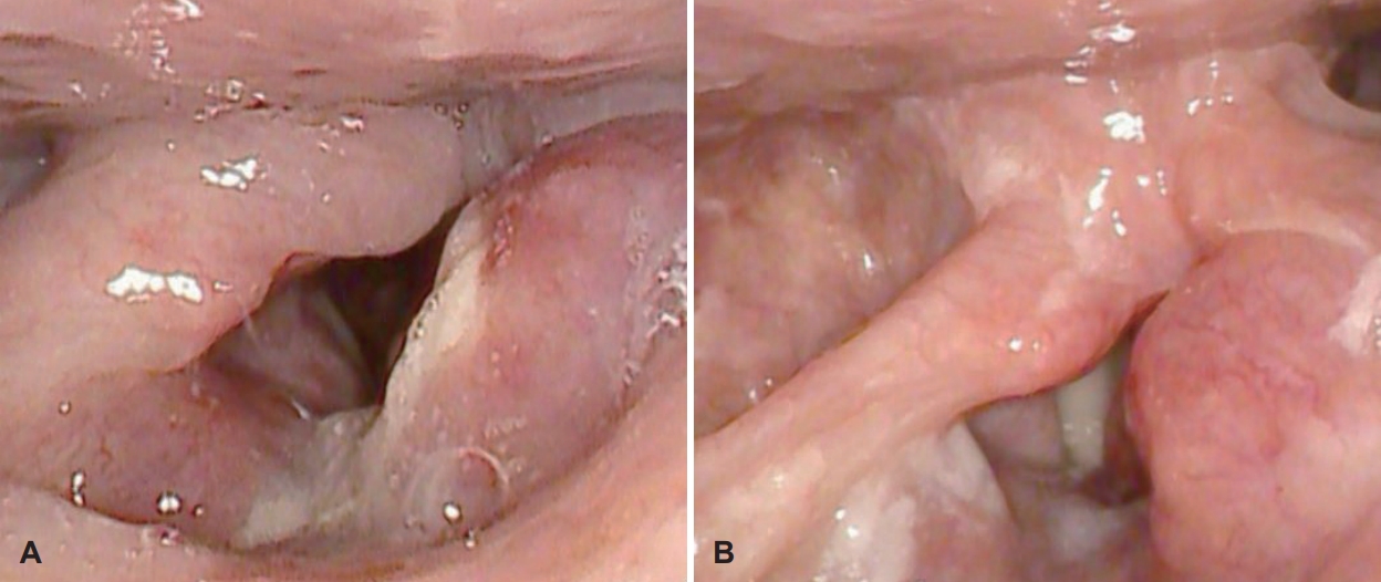
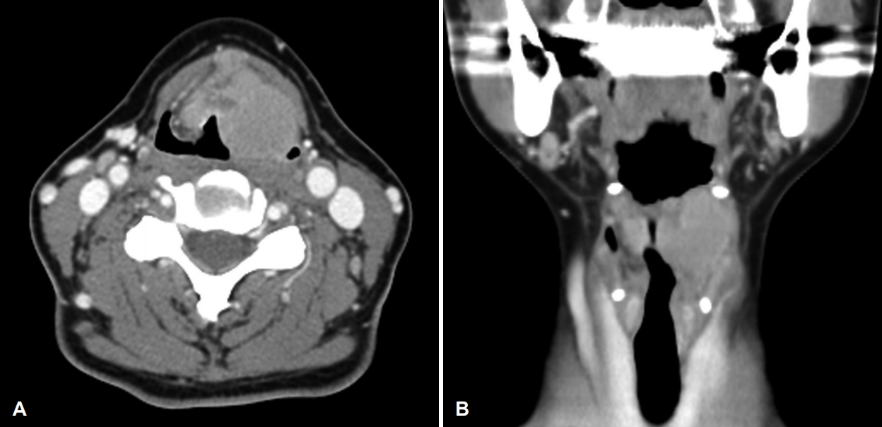
Flexible laryngoscopic examination revealed a mass-like lesion in the left supraglottic area, and the left vocal cord mobility decreased slightly (Fig. 1). Contrast-enhanced CT showed a 3×3-cm mass in the supraglottic region (Fig. 2). The mass involved the false vocal cord, pre-epiglottic space, both aryepiglottic folds, and paraglottic space.
Laryngeal microscopic surgery (LMS) for biopsy was performed under general anesthesia to rule out laryngeal cancer. The mucosa overlying the supraglottic mass was relatively intact, and a punch biopsy was performed including the mucosa and parenchymal tissue. The tissue sample was sent to the pathology department for a frozen biopsy, which showed a benign lesion without evidence of malignancy.
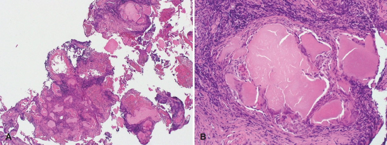
The postoperative course was uneventful, and there were no perioperative complications. Two weeks after the surgery, the biopsy result was confirmed as a rheumatoid nodule. The histopathology showed eosinophilic amorphous material scattered in the granulation tissue. Lymphocytic infiltration and fibrosis were also observed, and histiocytes and multinucleated giant cells surrounded the amorphous material (Fig. 3).
No further surgical interventions were performed for the laryngeal lesions, and the patient is being followed up at the outpatient clinic while taking medications for rheumatoid arthritis including MTX, hydroxychloroquine, and low dose steroid therapy. In the 6-month follow-up visit, the supraglottic mass was stable without any increase in size.
Discussion
The extra-articular manifestations of rheumatoid arthritis, such as rheumatoid nodules and rheumatoid vasculitis, may cause a variety of ocular, pulmonary, gastrointestinal, cardiovascular, renal, neurological, and hematological symptoms rather than locomotor disabilities [7].
The prevalence of laryngeal involvement in rheumatoid arthritis ranges from 0.3% to 12% [2,8-11]. The most common laryngeal manifestation of rheumatoid arthritis is the involvement of the cricoarytenoid joint, occurring in about 66% of laryngeal involvement cases, which may cause cricoarytenoid joint fixation and airway obstruction [9]. Other symptoms may include laryngeal mucosal edema, myositis of the intrinsic laryngeal muscles, inflammatory masses or rheumatoid nodules, and bamboo nodes [6]. The clinical presentation of laryngeal rheumatoid nodules vary from being asymptomatic to causing life-threatening respiratory symptoms and include sore throat, cough, foreign body sensation, odynophagia, hoarseness, dysphonia, change in voice quality, and respiratory distress [5,6]. The previous papers published in Korea have reported only two cases of vocal cord paralysis with cricoarytenoid joint fixation caused by rheumatoid arthritis [4,11]. To the best of our knowledge, this is the first case of rheumatoid nodules involving the larynx with voice change in Korea. In our case, the cause of decreased mobility of left vocal cord was due to the mass effect of rheumatoid nodule rather than cricoarytenoid joint fixation.
The diagnosis of laryngeal rheumatoid nodules needs a high degree of suspicion. CT, MRI, and laryngoscopic examination are useful for an early diagnosis. The final diagnosis can be made with histopathological examination [7]. In a case of laryngeal mass with rheumatoid arthritis, the possibility of rheumatoid nodules should be considered, and laryngeal cancer should be included in the differential diagnosis.
Laryngeal manifestations of rheumatoid nodules are rather benign, but sometimes they can be life-threatening. The treatment of rheumatoid nodules can be medical or surgical. In the early stages of the disease, medical treatment alone can be effective. The first-line medical treatment is the administration of steroids or nonsteroidal anti-inflammatory drugs to prevent the formation of nodules and fibrosis [12]. The steroids can be given systemically or locally for its anti-inflammatory effects.
MTX, which is widely used in the treatment of rheumatoid arthritis, may increase the size and number of rheumatoid nodules [13]. Therefore, if MTX is suspected to have caused the formation of rheumatoid nodules, discontinuing it can be considered. However, stopping MTX therapy is governed by several factors such as disease activity, drug availability, and cost concerns [14].
If the laryngeal rheumatoid nodules present with phonatory symptoms, voice therapy, steroid injection into the laryngeal mass or surgical intervention may be considered. However, in the presence of airway symptoms, surgical excision is necessary. In that case, LMS is performed under general anesthesia to remove the mass while preserving the overlying mucosa. In chronic cases where rheumatoid nodules invade the cricoarytenoid joint causing bilateral vocal cord fixation, an emergency tracheostomy is required [15].
In this study, the main symptom of patient is voice change, and there are no symptoms such as dyspnea and dysphagia. During 6 months of follow-up after diagnosis, the size of laryngeal rheumatoid nodule did not increase. Therefore, she has continued to take medication for rheumatoid arthritis rather than laryngeal surgery. However, if the size of rheumatoid nodule enlarges to cause dyspnea, stopping MTX or surgical excision will be necessary.
In conclusion, in rheumatoid arthritis patients with laryngeal mass, the possibility of rheumatoid nodules as well as laryngeal malignancy should be considered in the differential diagnosis of a laryngeal mass.