Introduction
Choristoma, also known as a hairy polyp, is commonly located in the nasopharynx or oropharynx of the head and neck region [1]. Choristoma is defined as an aggregation of mature polygerminal tissues that can be found anywhere in the body and is typically seen as a pedunculated polyp in children. It consists of ectoderm and mesoderm and is referred to as a bigerminal cell layer composition, which is often present in dermoids. In addition, its histopathological contents include sebaceous glands, cartilage, muscle fibers, hairy follicles. However, it remains controversial whether choristoma is a teratoma, which forms due to a congenital defect in the developmental process, or a neoplasm. Therefore, hairly polyp is similar to dermoid, but it is also considered to be more related to teratoma because it is differentiated by neoplasia recently.
Choristoma, also known as a hairy polyp that arises in the nasal cavity or nasopharynx, has rarely been reported in adults but is relatively common in children. Fewer than 10 cases have been described in adults [2-6]. Clinically, choristoma can present as a large symptomatic mass, but it often is hidden and more difficult to locate if it is small in size, especially in adults [2]. Symptoms that occur in adults due to choristoma include otalgia, hearing loss, recurrent epistaxis, and difficulty swallowing or with respiration.
Here, we report a choristoma detected along the nasopharyngeal wall that was causing nasal stuffiness and intermittent ear fullness in an adult. We also report for the first time the results of MRI of a nasopharyngeal choristoma in an adult, comparing with another benign mass in domestic journal. We expect our report to be educationally valuable for differentiating nasopharyngeal choristoma from other types of nasopharyngeal mass.
Approval from the Institutional Review Board was not required because this study did not include any human or animal studies (exemption number VC21ZISI0190 by the Institutional Review Committee, College of Medicine, the Catholic University, Korea).
Case
A 56-year-old male patient visited our outpatient clinic due to frequent nasal congestion, crust from the nasal cavity, and intermittent left ear fullness for the past six months. The patient reported exacerbating nasal stuffiness, especially when prone. He also complained of right-sided hearing loss and tinnitus since childhood. Pure tone analysis revealed normal left-sided hearing but a 50 dB air bone gap in the right ear. A narrow external auditory canal and tympanic membrane perforation on the right side were also observed.
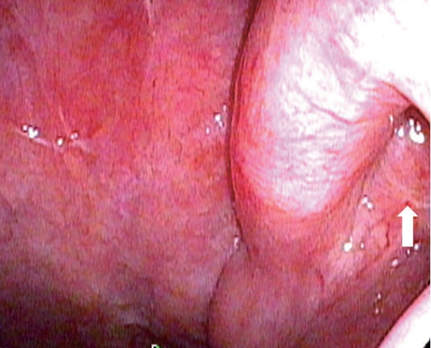
Upon endoscopy, a large polypoid mass with a stalk located on the lateral nasopharyngeal wall in front of the left rosenmuller fossa was detected (Fig. 1). The mass was located near the left torus tubarius and was obstructing the left eustachian tube orifice.
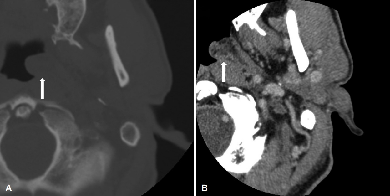
Paranasal sinus (PNS) CT scanning results showed a 27√Ч 13-mm-sized, heterogeneously enhanced, mass-like lesion in front of the left rosenmuller fossa (Fig. 2). No infiltration or bony osteolysis was observed, and no osteolytic findings were seen in the petrous temporal bone.
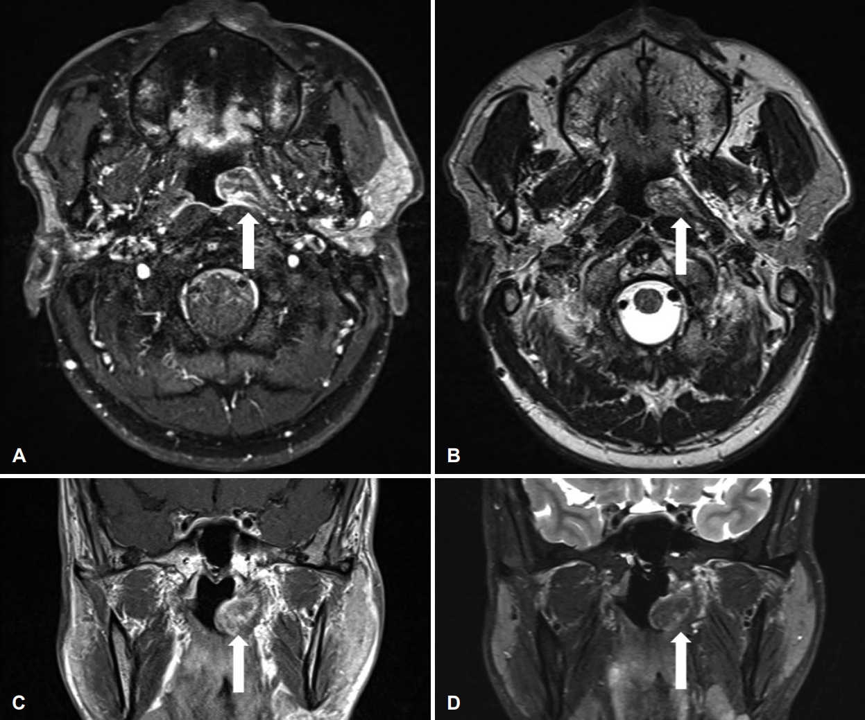
Based on the results of PNS MRI, the mass was dislocating the left torus tubarius muscle backward and spreading into the left eustachian tube opening (Fig. 3). The mass showed high signal intensity with mild rim enhancement on T1 weighted image and heterogenous signal intensity on T2 weighted image. From T2 weighted image, bright signal intensity showed low signal intensity with fat suppression. In addition, there was no left parapharyngeal space infiltration or preverbal muscle invasion. The function of the left eustachian tube was relatively well maintained. Diffusion restriction could not be detected due to artifacts.
The biopsy was performed at our outpatient clinic. The pathologic results suggested a benign polypoid lesion with squalid epithelium and inflammatory stroma. We attempted to distinguish between squamous cell papilloma, fibroepithelial polyp, and non-neoplastic stromal infiltration. The final pathological diagnosis was uncertain, so the patient underwent biopsy and resection with endoscopy under general anesthesia. The polypoid mass was located on the left local nasopharyngeal wall and was strongly attached to the eustachian tube wall. Completely removing the mass likely would have led to complications, such as eustachian tube stricture, so only part of the mass was removed. Further treatment was planned after confirmation of final diagnosis.
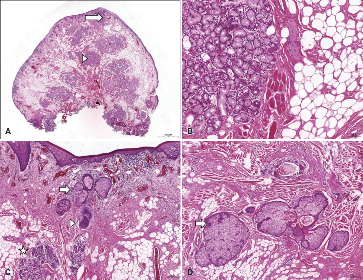
The pathological results showed mixed variable tissue with polypoid lesions and skin adnexal components (Fig. 4). The final pathologic diagnosis was choristoma or hairy polyp. Necrosis or mitotic activity was not observed.
The patient did not complain of nasal stuffiness, ear fullness, or recurrence postoperatively. Therefore, adjuvant therapy for the benign mass was not performed. No recurrence of the mass was noted during 1 year of postoperative followup period (Fig. 5).
Discussion
Most common nasopharyngeal masses in adults are malignant lesion. The most common malignant nasopharyngeal neoplasm in adults is the nasopharyngeal carcinoma. It also should be differential diagnosed with other mass such as lymphoma, tuberculosis, sarcoidosis, Wegner granulomatosis and angiofibroma [7]. Unlike adults, most nasopharyngeal masses in pediatrics are benign. However, it is important to identify malignancies. Masses commonly identified in the pediatric nasopharyngeal region include adenoid hypertrophy, teratoma, ThornwaldtвАЩs cyst, angiofibroma, fibroma, chondroma, hemangioma, and choristoma [8].
Choristoma, also known as a hairy polyp, is composed of aggregated tissue and is often found in children (especially newborns) but rarely seen in adults. Therefore, the developmental mechanism is considered to be a congenital problem of the brachial arch or a problem in development of stem cells due to trauma, rather than a congenital defect [1]. There is also a corresponding view of neoplasm. An abnormality noted in formation of the pharyngeal arch would explain the problems connected to the eustachian tube [6]. Since this type of mass is pathologically composed of mesoderm and ectoderm, choristoma has been described as both a dermoid and a teratoma, bigeminal mature teratoma, or benign teratoma [1,6].
Main pathologic differential diagnosis includes mixed epithelial and mesenchymal hamartoma and teratoma. Mixed epithelial and mesenchymal hamartoma shows seromucinous glandular epithelial proliferation along with the mesenchymal components, e.g., adipose tissue and skeletal muscle, similar to the choristoma. However, mixed epithelial and mesenchymal hamartomas are usually covered with respiratory-type surface epithelium and lack an adnexal component. According to the ArnoldвАЩs classification, teratoma contains endodermal derived tissue and a wide variety of tissue types [1]. The absence of these features that are usually seen in the teratoma allows for distinguishing choristoma from the teratoma.
Nasopharyngeal or oropharyngeal choristoma in children and adults can be identified using an endoscope. Most reported choristomas in adults are located on the lateral wall of the nasopharynx and sometimes on the palatine arches or lower lips. The incidence rate is high in female children, but the frequency in adults is unknown.
Prior to histological confirmation, it is also important to make a differential diagnosis with other tumors of the nasal cavity or nasopharynx through non-invasive imaging screening such as CT or MRI radiologic findings. In the presented case, the mass was high signal intensity on T1 weighted imaging with mild rim enhancement. On the T2 weighted image, it showed heterogenous signal intensity and bright signal intensity portion changed to low signal intensity with fat suppression. These characteristics in combination with other findings may help distinguish choristoma from other benign masses of the nasal cavity of the nasopharynx. Differential diagnosis using MRI for other benign masses of the nasal cavity or nasopharynx was summarized in Table 1. From Table 1, dermoid and angiomatous polyp show high signal intensity on T1 weighted image. However, angiomatous polyp shows more marked enhancement on T1 weighted image and higher signal intensity on T2 weighted image than choristoma. The MRI finding of previously reported dermoid was high signal intensity on T1 weighted image and variable intensity on T2 weighted image. This difference might due to the amount of adipose tissue.
Because the cost-effectiveness of MRI in nasopharyngeal mass screening is not relatively high, it should be used as the useful diagnostic tool for nasopharyngeal choristoma. MRI has no radiation exposure and can clearly demonstrate characteristics of the mass and its relationship with surrounding tissues and vessels. It also helps to find out the surgical plan.
We also found out that the MRI findings of choriostoma in adults are different from in neonates and children as reported in other papers [15]. Unlike the MRI finding of adults described earlier, choristoma in neonates and children show high or mixed signal intensity on T2 weighted image. This might be because the extent of adipose tissue or other components in choristoma is different.
Treatment of benign tumors in the nasal cavity or nasopharynx typically involves surgical resection, but the approach should consider the pathology results and structure in the nasal cavity or nasopharynx. If choristoma is suspected, treatment should be carried out via endoscopic excision or transoral excision.
In this case report, we described the clinical characteristics and MRI findings of a choristoma in the nasopharynx. We suggest that differential diagnosis include MRI findings of other benign masses of the nasal cavity or nasopharynx through a review of the literature.