м„ң лЎ
Primitive myxoid mesenchymal tumor of infancy (PMMTI)лҠ” 2006л…„ Alaggio л“ұ[1]мқҙ мІҳмқҢмңјлЎң л°ңн‘ңн•ң мҶҢм•„мқҳ м—°мЎ°м§Ғ(soft tissue)м—җ л°ңмғқн•ҳлҠ” л§Өмҡ° л“ңл¬ё м•…м„ұмў…м–‘мқҙлӢӨ. мқҙ м•…м„ұмў…м–‘мқҖ мӢ мғқм•„л¶Җн„° л°ңкІ¬лҗ мҲҳ мһҲмңјл©° мЈјлЎң 1м„ёк°Җ лҗҳкё° м „м—җ мІҙк°„, мӮ¬м§Җ, л‘җкІҪл¶Җ л“ұ лӢӨм–‘н•ң мҳҒм—ӯм—җм„ң л°ңкІ¬лҗңлӢӨ. көӯмҶҢм ҒмңјлЎң л§Өмҡ° кіөкІ©м Ғмқё м§ҲнҷҳмңјлЎң м•Ңл Өм ё мһҲмңјлӮҳ м•„м§Ғ к·ё мӮ¬лЎҖк°Җ л“ңл¬јм–ҙ мһҗм„ён•ң мһ„мғҒкІҪкіјлҘј м•Ңкё° нһҳл“ӨлӢӨ[2]. нҳ„мһ¬к№Ңм§Җмқҳ м№ҳлЈҢл°©лІ•мңјлЎңлҠ” мҲҳмҲ м Ғ м№ҳлЈҢлҘј нҶөн•ҳм—¬ мҷ„м „ м Ҳм ңлҘј н•ҳлҠ” кІғмқҙ мӣҗм№ҷмқҙлӮҳ, м Ҳм ңн•ҳлҚ”лқјлҸ„ лі‘ліҖмқҳ мһ¬л°ң л°Ҹ нғҖ л¶Җмң„лЎң м „мқҙк°Җ кҙҖм°°лҗ мҲҳ мһҲлӢӨ[3]. м Ҳм ңлҘј н• мҲҳ м—ҶлҠ” кІҪмҡ°м—җлҠ” н•ӯм•”мҡ”лІ•, л°©мӮ¬м„ мҡ”лІ• л“ұмқҳ м№ҳлЈҢлҘј мӢңлҸ„н• мҲҳ мһҲмңјлӮҳ, мқҙл§Ҳм ҖлҸ„ м Ҳл°ҳ м •лҸ„м—җм„ңлҠ” м№ҳлЈҢм—җ мӢӨнҢЁн•ңлӢӨ[4]. м Җмһҗл“ӨмқҖ мҡ°м—°нһҲ л°ңкІ¬лҗң нӣ„л‘җл¶Җ(occipital area)мқҳ мў…л¬јмқ„ мЈјмҶҢлЎң лӮҙмӣҗн•ң 3м„ё м—¬м•„ PMMTI мӮ¬лЎҖлҘј кІҪн—ҳн•ҳмҳҖкё°м—җ мқҙм—җ лҢҖн•ң мһ„мғҒ кІҪкіј л°Ҹ м№ҳлЈҢ кІ°кіјм—җ лҢҖн•ҙ ліҙкі н•ҳкі мһҗ н•ңлӢӨ.
мҰқ лЎҖ
3м„ё м—¬м•„к°Җ 11к°ңмӣ” м „ мҡ°м—°нһҲ л§Ңм ём§„ мҡ°мёЎ нӣ„л‘җл¶Җмқҳ мў…л¬јмқ„ мЈјмҶҢлЎң лӮҙмӣҗн•ҳмҳҖлӢӨ(Fig. 1). мў…л¬јмқҙ м җм җ м»Өм§ҖлҠ” м–‘мғҒмңјлЎң л°ңкІ¬ 1к°ңмӣ”м§ём—җ нғҖ лі‘мӣҗм—җ лӮҙмӣҗн•ҳм—¬ мӢңн–үн•ң мҙҲмқҢнҢҢмғҒ мҡ°мёЎ нӣ„л‘җл¶Җм—җм„ң 2.8Г—2.3Г—1.8 cmмқҳ лӮӯм„ұ ліҖнҷ”(cystic change)к°Җ мһҲлҠ” мў…м–‘м„ұ лі‘ліҖмқҙ кҙҖм°°лҗҳм–ҙ м„ём№ЁнқЎмқёкІҖмӮ¬(fine needle aspiration)лҘј мӢңн–үн•ҳмҳҖмңјлӮҳ, 비진лӢЁ мҶҢкІ¬(non-diagnostic)мңјлЎң кІҪкіј кҙҖм°° н•ҳмҳҖлӢӨ. л°ңкІ¬ 6к°ңмӣ”м§ё мһ¬лӮҙмӣҗн•ҳм—¬ мӢңн–үн•ң мҙҲмқҢнҢҢмғҒ 3.1Г—3.8 cmмқҳ л№„к· м§Ҳнҳ• лҸҷм—җмҪ” мў…м–‘ мҶҢкІ¬(heterogeneous isoechoic mass)мңјлЎң нҒ¬кё°к°Җ мҰқк°Җн–Ҳкі кІҪл¶Җ мһҗкё°кіөлӘ…мҳҒмғҒ(MRI)мғҒм—җм„ңлҠ” мҡ°мёЎ нӣ„л‘җл¶Җм—җ 2.7Г—2.8Г—3.8 cmмқҳ T1 кі мӢ нҳёк°•лҸ„(high signal), T2 л№„к· м§Ҳ кі мӢ нҳёк°•лҸ„(heterogeneous high signal) мҶҢкІ¬мңјлЎң м—°мЎ°м§Ғ мңЎмў…(soft tissue sarcoma)мқҙ мқҳмӢ¬лҗҳм§Җл§Ң л‘җк°ңм Җмқҳ лјҲ м№ЁлІ”мқҖ кҙҖм°°лҗҳм§Җ м•Ҡм•ҳлӢӨ(Fig. 2A). л°ңкІ¬ 10к°ңмӣ”м§ё нғҖ лі‘мӣҗм—җм„ң м Ҳм ң мғқкІҖмқ„ кі„нҡҚн•ҳмҳҖмңјлӮҳ нҷҳмһҗ мӮ¬м •мғҒ мӢңн–үн•ҳм§Җ м•Ҡкі мҙҲмқҢнҢҢ мң лҸ„н•ҳ мӨ‘мӢ¬л¶Җл°”лҠҳмғқкІҖ(core needle biopsy)лҘј мӢңн–үн•ҳмҳҖмңјл©°, м•…м„ұлҸ„к°Җ нҷ•мӢӨн•ҳм§Җ м•ҠмқҖ мӨ‘к°„м—Ҫ мў…м–‘(mesenchymal tumor) мҶҢкІ¬мқ„ ліҙм—¬ 추к°Җм Ғмқё нҸүк°Җ л°Ҹ м№ҳлЈҢлҘј мң„н•ҙ ліё кё°кҙҖмңјлЎң мқҳлў°лҗҳм—ҲлӢӨ.
мў…л¬ј л°ңкІ¬ 11к°ңмӣ”м§ёк°Җ лҗҳлҠ” ліёмӣҗ лӮҙмӣҗ лӢ№мӢң мҡ°мёЎ нӣ„л‘җл¶Җм—җ м•Ҫ 4 cm нҒ¬кё°мқҳ лӢЁлӢЁн•ң л¬ҙнҶөмқҳ мў…л¬јмқҙ мҙү진лҗҳм—Ҳмңјл©°, кІҪл¶Җ м»ҙн“Ён„°лӢЁмёөмҙ¬мҳҒ(CT)мғҒ м•Ҫ 3.8Г—3.0Г—3.7 cmмқҳ л№„к· м§Ҳм„ұмқҳ мЎ°мҳҒ мҰқк°•мқҙ лҡңл ·н•ң мў…м–‘(heterogeneous and avidly enhancing mass)мқҙ мҡ°мёЎ нӣ„л‘җл¶Җм—җм„ң кҙҖм°°лҗҳл©° мЈјліҖл¶Җмқҳ лјҲ м№ЁмӢқмқҙлӮҳ мЈјліҖ м§Җл°© м№ЁмңӨ(perilesional fat infiltration)мқҖ кҙҖм°°лҗҳм§Җ м•Ҡм•ҳлӢӨ(Fig. 2B). мҙҲмқҢнҢҢмғҒм—җм„ңлҠ” мҡ°мёЎ нӣ„л‘җл¶Җм—җ м•Ҫ 4.0Г—2.7 cm нҒ¬кё°мқҳ лӢЁлӢЁн•ң мў…м–‘мқҙ кҙҖм°°лҗҳл©° лі‘ліҖмқҳ мЈјмң„лЎң нҳҲкҙҖ 분нҸ¬(peripheral vascularity)к°Җ ліҙмҳҖлӢӨ(Fig. 2C and D). мў…м–‘мқҳ к°•м§ҒлҸ„(stiffness)лҘј м•Ңм•„ліҙкё° мң„н•ҳм—¬ мӢңн–үн•ң ліҖнҳ• нғ„м„ұ мҙҲмқҢнҢҢ(strain elastography)мғҒ мў…м–‘мқҳ мғҒлҢҖм Ғмқё ліҖнҳ•лҘ (strain ratio)мқҖ нҸүк· 6.4лЎң н”јн•ҳ л°Ҹ л“ұм„ёлӘЁк·ј к·јмңЎкіј 비көҗн•ҳмҳҖмқ„ л•Ң м•Ҫ 6.4л°° лӢЁлӢЁн•ң мҶҢкІ¬мқ„ ліҙмҳҖлӢӨ(Fig. 2E). мЎ°мҳҒмҰқк°•мҙҲмқҢнҢҢ(contrast enhanced US)м—җм„ңлҠ” лі‘ліҖмқҳ н‘ңмёө(superficial portion)м—җм„ң кҙҖлҘҳ кІ°мҶҗ(perfusion defect)мқҙ ліҙм—¬ лӮҙл¶Җ кҙҙмӮ¬(internal necrosis) нҳ№мқҖ м җм•Ўм„ұ мҡ”мҶҢ(myxoid component)к°Җ мһҲмқ„ кІғмңјлЎң нҢҗлӢЁлҗҳм—ҲлӢӨ(Fig. 2F). ліё кё°кҙҖм—җм„ң лӢӨмӢң н•ңлІҲ мӢңн–үн•ң мҙҲмқҢнҢҢ мң лҸ„н•ҳ мӨ‘мӢ¬л¶Җл°”лҠҳмғқкІҖмғҒ PMMTIм—җ к°ҖмһҘ к·јм ‘н•ҳлӮҳ мҳҒм•„ 섬мң мңЎмў…(infantile fibrosarcoma)мқ„ л°°м ңн• мҲҳ м—ҶлӢӨлҠ” лі‘лҰ¬ кІ°кіјлҘј нҷ•мқё нӣ„ мҲҳмҲ м Ғ м Ҳм ңлҘј кі„нҡҚн•ҳмҳҖлӢӨ.
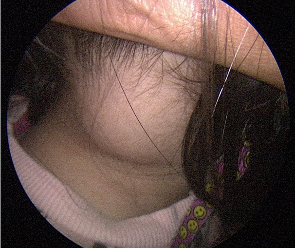
мҲҳмҲ мӢӨм—җм„ң нҷ•мқё лӢ№мӢң мҡ°мёЎ нӣ„л‘җл¶Җм—җ м•Ҫ 4 cm нҒ¬кё°мқҳ мў…л¬јмқҙ л“ұм„ёлӘЁк·јліҙлӢӨ к№ҠкІҢ мң„м№ҳн•ҳкі мһҲм—Ҳкі л“ұм„ёлӘЁк·јкіј мң м°©мқҙ мһҲм–ҙ мқјл¶Җ к·јмңЎмқ„ нҸ¬н•Ён•ҳм—¬ мў…л¬јмқ„ л°•лҰ¬н•ҳмҳҖлӢӨ. мҲҳмҲ лӢ№мӢң мңЎм•ҲмғҒ мў…л¬јмқҙ мЈјмң„ мЎ°м§Ғкіј мң м°©мқҙ мһҲм–ҙ ліҙмҳҖкё° л•Ңл¬ём—җ мЈјмң„ мЎ°м§Ғмқ„ нҸ¬н•Ён•ҳм—¬ мў…л¬јмқ„ нҢҢм—ҙ м—Ҷмқҙ м ңкұ°н•ҳмҳҖлӢӨ(Fig. 3A).
мҲ нӣ„ мЎ°м§ҒкІҖмӮ¬ кІ°кіјмғҒ PMMTIк°Җ 진лӢЁлҗҳм—Ҳкі мў…л¬јмқҳ нҒ¬кё°лҠ” 3.7Г—3.2Г—2.2 cmмңјлЎң мёЎм •лҗҳм—ҲлӢӨ. лі‘лҰ¬ кІҖмӮ¬мғҒ 100л°° л°°мңЁм—җм„ң мў…м–‘мқҳ лӢӨм–‘н•ң л¶Җ분м—җм„ң нҳҲкҙҖмЈјмң„м„ёнҸ¬мў… м–‘мғҒмқҳ нҳҲкҙҖ(hemangiopericytoma-like vessel)мқҙ кҙҖм°°лҗҳм—ҲлӢӨ(Fig. 3B). 400л°° л°°мңЁм—җм„ң нғҖмӣҗнҳ• лҳҗлҠ” мӣҗнҳ•мқҳ н•ө л°Ҹ мҲҳнҸ¬м„ұ м—јмғүм§Ҳ нҳ•нғң(vesicular chromatic pattern)к°Җ ліҙмқҙл©° м—°н•ң нҳёмӮ°м„ұ мў…м–‘м„ёнҸ¬м§Ҳ(pale eosinophilic tumor cytoplasm)мқ„ ліҙмқёлӢӨ(Fig. 3C and D). мў…л¬јмқ„ нҢҢм—ҙ м—Ҷмқҙ м ңкұ°н•ҳмҳҖкё° л•Ңл¬ём—җ лі‘лҰ¬ кІ°кіјмғҒ м Ҳм ңм—° мқҢм„ұ(margin negative)мқҙ лӮҳмҳ¬ кІғмңјлЎң кё°лҢҖн•ҳмҳҖмңјлӮҳ м Ҳм ңм—° м–‘м„ұ(margin positive) мҶҢкІ¬мқҙ лӮҳмҷ”кі , ліҙнҳёмһҗм—җкІҢ мһ¬мҲҳмҲ н•„мҡ”м„ұ м„ӨлӘ…н•ҳмҳҖмңјлӮҳ мЈјліҖ кө¬мЎ°л¬јмқҳ кҙ‘лІ”мң„н•ң м Ҳм ңлҘј лӢ№мһҘ мӣҗн•ҳм§Җ м•Ҡкі м¶”м Ғ кҙҖм°°мқ„ мӣҗн•ҳм—¬ мҶҢм•„ нҳҲм•Ўмў…м–‘лӮҙкіјмҷҖ мғҒмқҳн•ҳм—¬ лӢЁкё°к°„ мҳҒмғҒ 추м Ғ кҙҖм°°н•ҳл©ҙм„ң н•„мҡ” мӢң мһ¬мҲҳмҲ л°Ҹ 추к°Җ н•ӯм•”м№ҳлЈҢлҘј кі л Өн•ҳкё°лЎң н•ҳмҳҖлӢӨ. мҲ нӣ„ 1к°ңмӣ”м—җ мӢңн–үн•ң MRIмғҒ мҲҳмҲ л¶Җмң„лЎң м•ЎмІҙ м ҖлҘҳ л°Ҹ мЎ°мҳҒ мҰқк°•лҗҳлҠ” лі‘ліҖмқҙ кҙҖм°°лҗҳм—ҲмңјлӮҳ(Fig. 2G), мҲҳмҲ 3к°ңмӣ”м—җ мӢңн–үн•ң MRI T1 к°•мЎ° мқҙлҜём§Җм—җм„ң н•ҙлӢ№ л¶Җмң„мқҳ м „мІҙм Ғмқё мЎ°мҳҒ мҰқк°•мқҙ к°җмҶҢн•ң мҶҢкІ¬мқ„ ліҙмқҙл©° лӘ…нҷ•н•ң мў…л¬ј м–‘мғҒмқҳ лі‘ліҖмқҙ ліҙмқҙм§Җ м•Ҡм•ҳлӢӨ. мҲҳмҲ 6к°ңмӣ” мӢңм җм—җлҸ„ мӢ мІҙ 진찰мғҒ мҡ°мёЎ нӣ„л‘җл¶Җм—җ мў…л¬ј л“ұ нҠ№мқҙмҶҢкІ¬мқ„ ліҙмқҙм§Җ м•Ҡм•„ кІҪкіјлҘј м§Җмјңліј мҳҲм •мқҙлӢӨ(Fig. 2H).
кі м°°
PMMTIлҠ” кё°мЎҙк№Ңм§Җ лҜёл¶„нҷ” мңЎмў…(undifferentiated sarcoma)мңјлЎң 분лҘҳлҗҳкұ°лӮҳ м„ мІңм„ұ мҳҒм•„ 섬мң мңЎмў…мңјлЎң 분лҘҳлҗҳм—ҲлӢӨ. н•ҳм§Җл§Ң л‘җ мңЎмў…кіјлҠ” лӢӨлҘҙкІҢ мӣҗмӢң лҜём„ұмҲҷ мӨ‘к°„м—Ҫ м„ёнҸ¬(primitive mesenchymal cell)к°Җ м„ёнҸ¬н•ҷм Ғ мқҙнҳ•м„ұмҰқмқ„ к°–лҠ” м җм•ЎмІҙ(distinctive myxoid background) лӮҙм—җм„ң мһҗлһҖлӢӨлҠ” нҠ№м§•мқ„ к°Җм§Җл©° лҚ” кіөкІ©м Ғмқё мһ„мғҒ кІҪкіјлҘј лӮҳнғҖлӮёлӢӨлҠ” м җмңјлЎң 2006л…„ Alaggio л“ұ[1]мқҙ л°ңн‘ңн•ң л…јл¬ём—җм„ң PMMTIлқјлҠ” мҡ©м–ҙк°Җ мғҲлЎӯкІҢ мӮ¬мҡ©лҗҳм—ҲлӢӨ. PMMTIмқҳ 진лӢЁ кё°мӨҖмқҖ м•„м§Ғ м •н•ҙм§Җм§Җ м•Ҡм•ҳмңјлӮҳ, кё°мЎҙм—җ ліҙкі лҗң мӮ¬лЎҖл“Өмқҳ нҳ„лҜёкІҪ мҶҢкІ¬мқ„ мў…н•©н•ҳл©ҙ мў…м–‘м„ёнҸ¬лҠ” мӣҗмӢң 방추нҳ•(primitive spindle), лӢӨк°Ғнҳ•(polygonal) лҳҗлҠ” мӣҗнҳ•мқҳ лӘЁм–‘мқ„ к°Җм§Ҳ мҲҳ мһҲмңјл©° мў…м–‘м„ёнҸ¬мқҳ н•өмқҖ лӢЁмЎ°лЎңмҡ°л©ҙм„ң лҸҷмқјн•ң нҳ•нғңлҘј ліҙмқҙл©° н•өмқҳ м—јмғүм§ҲмқҖ к· мқјн•ҳкі н•өмҶҢмІҙлҠ” кұ°мқҳ кҙҖм°°лҗҳм§Җ м•ҠлҠ”лӢӨ. мқҙлҹ¬н•ң мў…м–‘м„ёнҸ¬л“ӨмқҖ м җм•Ўм„ұ кё°м§Ҳ(myxoid stroma)мқ„ кё°л°ҳмңјлЎң м„ұмһҘн•ҳл©° кі м„ёнҸ¬м¶©мӢӨлҸ„мҷҖ м Җм„ёнҸ¬м¶©мӢӨлҸ„лҘј лӘЁл‘җ ліҙмқҙлҠ” кІҪмҡ°к°Җ л§ҺлӢӨ. мў…м–‘м„ёнҸ¬ лҚ©м–ҙлҰ¬ мӮ¬мқҙлЎң нҳҲкҙҖ мҰқмӢқмқҙ л§Һмңјл©° нҳҲкҙҖмЈјмң„м„ёнҸ¬мў… м–‘мғҒмқҳ нҳҲкҙҖ лӘЁм–‘лҸ„ мһҗмЈј кҙҖм°°лҗҳл©° м Җл°°мңЁмғҒм—җм„ң мў…м–‘мқҖ кІ°м Ҳнҳ•мңјлЎң мһҗлқјлҠ” кІҪмҡ°к°Җ л§Һмңјл©°, к°Ғк°Ғмқҳ кІ°м Ҳл“ӨмқҖ мҪңлқјкІҗ кё°м§ҲлЎң кө¬л¶„лҗңлӢӨ.
PMMTIмқҳ м№ҳлЈҢлҠ” м•„м§Ғ м •лҰҪлҗҳм–ҙ мһҲм§Җ м•Ҡм§Җл§Ң 2020л…„ Asaftei л“ұ[4]мқҙ л°ңн‘ңн•ң л…јл¬ём—җ л”°лҘҙл©ҙ 2006л…„л¶Җн„° 2020л…„к№Ңм§Җ PMMTIлҠ” мҙқ 29мҳҲк°Җ л°ңн‘ңлҗҳм—Ҳмңјл©° мқҙ мӨ‘ м№ҳлЈҢ л°©лІ•мқ„ м•Ң мҲҳ м—ҶлҠ” 10кұҙмқ„ м ңмҷён•ҳл©ҙ 17мҳҲм—җм„ң мҲҳмҲ мқ„, 1мҳҲм—җм„ң н•ӯм•”м№ҳлЈҢлҘј, 1мҳҲм—җм„ң мҲҳмҲ кіј н•ӯм•”м№ҳлЈҢлҘј лі‘н–үн•ҳмҳҖлӢӨ. мқҙ мӨ‘ мҲҳмҲ нӣ„ кІ°кіјлҘј м•Ң мҲҳ м—ҶлҠ” 1кұҙмқ„ м ңмҷён•ң мҙқ 17мҳҲмқҳ мҲҳмҲ мӮ¬лЎҖ мӨ‘ 65% (11/17)м—җм„ңлҠ” мҲ нӣ„ мһ”м—¬ лі‘ліҖмқҙ мһҲм—Ҳмңјл©° 35% (6/17)м—җм„ңлҠ” мһ”м—¬ лі‘ліҖм—Ҷмқҙ м ңкұ°к°Җ мқҙлЈЁм–ҙмЎҢлӢӨ. мһ”м—¬ лі‘ліҖмқҙ мһҲлҠ” 11мҳҲ мӨ‘ 8мҳҲм—җм„ңлҠ” мһ¬л°ңн•ҳмҳҖмңјл©°(73%) мқҙ мӨ‘ 2кұҙм—җм„ңлҠ” 2м°Ё м№ҳлЈҢлЎң н•ӯм•”м№ҳлЈҢлҘј н•ҳмҳҖмңјлӮҳ мһ¬л°ңн•ҳм—¬ мҲҳмҲ м Ғ м№ҳлЈҢлҘј н•ҳмҳҖкі [1,5], 2кұҙм—җм„ңлҠ” 2м°Ё м№ҳлЈҢлЎң мҲҳмҲ кіј н•ӯм•” лі‘н•© мҡ”лІ•мқ„[1,6], 4кұҙм—җм„ңлҠ” мҲҳмҲ мқ„ н•ҳмҳҖлӢӨ[1,3,7,8]. мһ”м—¬ лі‘ліҖ м—Ҷмқҙ м ңкұ°к°Җ мқҙлЈЁм–ҙ진 6мҳҲм—җм„ңлҠ” 1кұҙл§Ңмқҙ мһ¬л°ңн•ҳмҳҖмңјл©°(17%) 2м°Ё м№ҳлЈҢлЎң мһ¬мҲҳмҲ мқ„ н•ҳмҳҖмңјлӮҳ лӢӨмӢң мһ¬л°ңн•ҳм—¬ м№ҳлЈҢ м—Ҷмқҙ кІҪкіј кҙҖм°° н•ҳмҳҖлӢӨ[3]. н•ӯм•”м№ҳлЈҢлҘј лӢЁлҸ…мңјлЎң мӢңн–үн•ң кІҪмҡ°лҠ” 2кұҙмқҳ мӮ¬лЎҖк°Җ мһҲм—Ҳмңјл©° Cuthbertson л“ұ[9]мқҖ 1м°Ё н•ӯм•”м№ҳлЈҢ нӣ„ мһ¬л°ңн•ҳм—¬ 2м°Ё м№ҳлЈҢлЎң мҲҳмҲ мқ„ н•ҳмҳҖмңјлӮҳ мһ¬л°ңн•ҳм—¬ 3м°Ё м№ҳлЈҢлЎң мҲҳмҲ л°Ҹ н•ӯм•”м№ҳлЈҢлҘј лі‘н–үн•ҳм—¬ мһ¬л°ңн•ҳм§Җ м•Ҡм•ҳкі Memmott л“ұ[10]мқҖ н•ӯм•”м№ҳлЈҢ нӣ„ лі‘ліҖмқҙ мһ¬л°ңн•ҳм§Җ м•Ҡм•ҳлӢӨкі ліҙкі н•ҳмҳҖлӢӨ.
PMMTIмқҳ мҲҳмҲ нӣ„ нҸүк· мһ¬л°ң кё°к°„мқҖ 4.4к°ңмӣ”(1-12к°ңмӣ”)лЎң л§Өмҡ° л№ лҘҙл©° мҲҳмҲ нӣ„ мһ”м—¬ лі‘ліҖмқҙ лӮЁм•„мһҲлҠ” кІҪмҡ°м—җлҸ„ лҚ” мқҙмғҒ нҒ¬кё° ліҖлҸҷ м—Ҷмқҙ м•Ҳм •м Ғмқё мғҒнғңлҘј мң м§Җн•ҳлҠ” кё°к°„мқҖ нҸүк· 25.3к°ңмӣ”(5-70к°ңмӣ”)мқҙлӢӨ[4]. мҡ°лҰ¬ мҰқлЎҖмқҳ кІҪмҡ° мҲ нӣ„ 1к°ңмӣ”м—җ мӢңн–үн•ң MRIмғҒ мҲҳмҲ л°”лӢҘм—җ м•ЎмІҙ м ҖлҘҳ л°Ҹ мЎ°мҳҒ мҰқк°• кҙҖм°°лҗҳм—ҲмңјлӮҳ мҲҳмҲ м—Ҷмқҙ кІҪкіј кҙҖм°° н•ҳмҳҖкі , мҲҳмҲ 3к°ңмӣ”м—җ мӢңн–үн•ң MRIмғҒ мҲҳмҲ 1к°ңмӣ”м—җ ліҙмқҙлҚҳ м•ЎмІҙ м ҖлҘҳлҠ” м—Ҷм–ҙмЎҢкі м „мІҙм Ғмқё мЎ°мҳҒ мҰқк°•мқҙ к°җмҶҢн•ң мҶҢкІ¬мқ„ ліҙмҳҖлӢӨ. н•ҳм§Җл§Ң нҸүк· мһ¬л°ң кё°к°„мқҙ 4.4к°ңмӣ”мһ„мқ„ к°җм•Ҳн•ҳкі 1л…„ л’Өм—җ мһ¬л°ңн•ң мӮ¬лЎҖлҸ„ мһҲлҠ” кІғмқ„ к°җм•Ҳн•ҳмҳҖмқ„ л•Ң, лӢЁкё°к°„ л°Ҹ мһҘкё°к°„ кІҪкіј кҙҖм°°мқҖ н•„мҲҳм Ғмқј кІғмңјлЎң ліҙмқёлӢӨ.
м•һм„ң м–ёкёүн•ң мҲҳмҲ нӣ„ мһ”м—¬ лі‘ліҖмқҙ ліҙмқҙм§Җ м•ҠлҠ” 6мҳҲ мӨ‘ мһ¬л°ңмқҙ л°ңмғқн•ҳм§Җ м•ҠмқҖ 5мҳҲмқҳ кіөнҶөм җмқҖ мҲҳмҲ нӣ„ лі‘лҰ¬ кІҖмӮ¬мғҒ нҳ„лҜёкІҪмғҒ мһ”м—¬ лі‘ліҖмқҳ мЎҙмһ¬к°Җ м—Ҷм—ҲлӢӨлҠ” м җмқҙлӢӨ[1,11-14]. мһ¬л°ңмқҙ л°ңмғқн•ң 1мҳҲм—җм„ңлҠ” кІҪл¶Җм—җ л°ңмғқн•ң 3 cmмқҳ лі‘ліҖмңјлЎң мҲҳмҲ нӣ„ мңЎм•Ҳм ҒмңјлЎңлҠ” мһ”м—¬ лі‘ліҖмқҙ м—Ҷм—ҲмңјлӮҳ нҳ„лҜёкІҪмғҒ мһ”м—¬ лі‘ліҖмқҳ мң л¬ҙлҘј нҷ•мқён•ҳм§Җ лӘ»н–ҲлҚҳ мӮ¬лЎҖмҳҖлӢӨ[3]. л”°лқјм„ң ліё мҰқлЎҖмқҳ кІҪмҡ°м—җлҸ„ 충분н•ң мҲҳмҲ м Ҳм ңм—°мқҳ нҷ•ліҙк°Җ лҗҳм§Җ м•Ҡм•ҳкё° л•Ңл¬ём—җ мӣҗм№ҷмғҒмңјлЎңлҠ” 추к°Җм Ғмқё м Ҳм ң л°Ҹ ліҙмЎ° л°©мӮ¬м„ мҡ”лІ• л“ұмқҳ м№ҳлЈҢлҘј н•ҳлҠ” кІғмқҙ мҳҲнӣ„к°Җ лҚ” мўӢмқ„ мҲҳ мһҲмңјлӮҳ, нҷҳмһҗ л°Ҹ ліҙнҳёмһҗмҷҖ мғҒмқҳн•ҳм—¬ л°Җм ‘н•ң кІҪкіј кҙҖм°°мқ„ мӢңн–үн•ҳкё°лЎң н•ҳмҳҖлӢӨ.
кІ°лЎ м ҒмңјлЎң PMMTIмқҙ м Ҳм ңк°Җ к°ҖлҠҘн•ң л¶Җмң„м—җ мһҲкі мІ« лІҲм§ё м№ҳлЈҢлЎң мҲҳмҲ м Ғ м№ҳлЈҢлҘј м„ нғқн–Ҳмқ„ кІҪмҡ° нҳ„лҜёкІҪмғҒ мһ”м—¬ лі‘ліҖмқҙ м—Ҷмқҙ м Ҳм ңн•ҳлҠ” кІғмқҙ мөңмғҒмқҳ м„ нғқмңјлЎң ліҙмқҙл©° мҲҳмҲ мӢң мў…л¬јмқ„ н”јл§үм—җ л¶ҷм—¬м„ң м Ҳм ңн•ҳлҠ” кІғліҙлӢӨлҠ” кІҪкі„лҘј 충분нһҲ м¶”кі м Ҳм ңлҘј н•ҳлҠ” кІғмқҙ мһ¬л°ңмқ„ мӨ„мқҙлҠ” лҚ° лҸ„мӣҖмқ„ мӨ„ мҲҳ мһҲмқ„ кІғмңјлЎң ліҙмқёлӢӨ.