Successful Treatment of Postauricular Muscle Myokymia Using Low-Dose Botulinum Toxin Type A
저용량 A형 보툴리눔 독소를 이용한 후이개근 근떨림증의 치료 1예
Article information

Abstract
Postauricular muscle myokymia is a rare focal movement disorder caused by involuntary activation of the posterior auricular muscle. We report a 44-year-old woman with a 2-year history of intermittent bursts of rapid, irregular twitching localized to the right postauricular region, which aggravated when lying on her right side or turning her head rightward. Oxcarbazepine was ineffective. In a single injection session, botulinum toxin type A (Nabota, 10 U in 0.2 mL; 50 U/mL) was administered into the posterior auricular muscle. Given the small size and superficial location of the muscle, a concentrated, low-volume formulation was chosen to confine the toxin to the target area. Symptoms markedly improved within 3 days and resolved completely by day 5. Visual Analogue Scale decreased from 5 to 0 at 2 weeks, remained 0 at 3 months, and was 1 at 6 months without requiring further treatment. No adverse effects occurred. This case illustrates that the botulinum toxin type A can be a safe and effective option for focal postauricular muscle myokymia when the symptomatic muscle is accurately identified.
Introduction
Involuntary auricular and peri-auricular movements are rare and may present as auricular myoclonus, ear dyskinesia, or focal movement disorders involving the extrinsic auricular muscles [1-3]. These muscles, including the posterior auricular muscle, are innervated by distal branches of the facial nerve and may exhibit abnormal activation when neural hyperexcitability or fascial tension is present. The posterior auricular muscle, in particular, has been implicated in posterior auricular dystonia, muscular tinnitus, and retroauricular twitching, although reports remain scarce [1,4,5]. Because of this rarity, the clinical course, optimal evaluation, and evidence-based treatment strategies remain inadequately defined.
Botulinum toxin type A has demonstrated therapeutic benefit across various auricular movement disorders, including ear-wiggling tics, muscular tinnitus, auricular myoclonus, and auricular dystonia [3-6]. However, previously reported doses vary widely—ranging from 5 U to 40 U—reflecting anatomical variability of extrinsic auricular muscles and challenges in accurately localizing the symptomatic focus [2-4]. Furthermore, diffusion to adjacent structures such as the sternocleidomastoid or occipitalis may lead to undesirable complications, emphasizing the importance of precise dosing and targeted injection [7].
Here, we describe a case of isolated postauricular muscle myokymia successfully treated with low-dose (10 U) botulinum toxin injection. This case highlights accurate clinical identification of the posterior auricular muscle as the symptomatic source and provides practical considerations for safe and effective injection in this anatomically compact region.
Case
A 44-year-old woman presented with a 2-year history of intermittent involuntary twitching over the right postauricular region. Symptoms were posture-dependent, worsening when she lay on her right side or rotated her head rightward. She reported intermittent fullness and mild tinnitus but denied otalgia, vertigo, autophony, or hearing loss. Her medical history included Hashimoto’s thyroiditis controlled with levothyroxine.
Otoscopy revealed a slightly widened external auditory canal but an intact tympanic membrane. Pure-tone audiometry and tympanometry were normal. Temporal bone CT showed no abnormalities, and laboratory results were unremarkable. On physical examination, intermittent bursts of rapid, irregular twitching were observed strictly within the right postauricular soft tissue without visible auricular movement, consistent with focal myokymia of the posterior auricular muscle. The contraction focus was clearly palpable along the expected course of the muscle (Fig. 1). The pre-treatment video documentation (Supplementary Video 1) was independently reviewed and confirmed by three senior otologists to ensure diagnostic accuracy and differentiate it from other facial movement disorders.
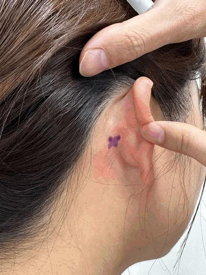
Clinical photograph showing the right postauricular region before injection. The suspected contraction focus was marked with a purple “X” over the posterior aspect of the auricle, corresponding to the clinically palpable area of involuntary contraction along the expected course of the posterior auricular muscle.
A trial of oxcarbazepine was initiated at a low starting dose of 75 mg twice daily (150 mg/day). No symptomatic improvement was observed during the 3-week trial, and as the patient preferred to pursue an alternative therapeutic approach, further dose escalation was not undertaken. Given the localized nature of the movement and failure of medical therapy, botulinum toxin injection was selected. Botulinum toxin type A (Nabota, Daewoong) was diluted to a concentration of 50 U/mL, and a total of 10 U was administered in a single injection session, delivering 5 U (0.1 mL) into the anterior portion of the posterior auricular muscle and 5 U (0.1 mL) into the posterior portion along its anatomical course (Fig. 2). Because the muscle is thin (1-2 mm) and superficial, a concentrated, low-volume formulation was selected in an attempt to confine the toxin to the target area. No adverse events occurred. Symptom improvement began on day 3 and was complete by day 5. The Visual Analogue Scale (VAS) score decreased from 5 to 0 at 2 weeks and remained 0 at 3 months. At 6 months, the patient reported only minimal symptom recurrence (VAS 1) that did not require further intervention.
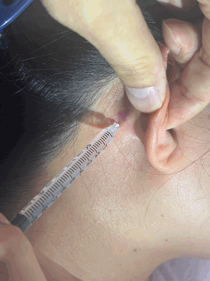
Botulinum toxin injection into the right postauricular region. Using a 1 cc insulin syringe, the needle was inserted perpendicularly at the marked site and redirected in multiple vectors to achieve fan-shaped infiltration without additional skin puncture. A total of 10 U was administered in two injections, delivering 5 U (0.1 mL) per point to the anterior and posterior portions of the posterior auricular muscle.
This case report was reviewed and approved by the Institutional Review Board of Pusan National University Hospital (IRB No. 2511-003-157). The requirement for written informed consent was waived because the report contains deidentified clinical information and poses minimal risk to the patient.
Discussion
Postauricular muscle myokymia is an uncommon focal movement disorder involving involuntary activation of the posterior auricular muscle, a thin and superficial muscle innervated by the posterior auricular branch of the facial nerve. The posterior auricular muscle lies among the extrinsic auricular muscles in close anatomical relation to adjacent periauricular structures (Fig. 3). Previous reports—including posterior auricular dystonia, auricular myoclonus, and moving ear syndrome—suggest that abnormal excitability of small extrinsic auricular muscles can generate visible twitching or dyskinetic movements in the peri-auricular region [1-3]. Unlike typical auricular myoclonus, which often produces rhythmic elevation or clicking tinnitus, postauricular myokymia may manifest as subtle wave-like contractions confined to retroauricular soft tissue. This specific condition is likely underreported because the postauricular region is frequently obscured by hair, and the focal muscle twitching may be overlooked or misclassified during routine clinical evaluations or dismissed as a benign variant of hemifacial spasm. The posture-dependent variability in this case, especially worsening during ipsilateral head rotation or dependent positioning, supports the hypothesis that mechanical tension along the mastoid-postauricular fascial plane can modulate excitability of distal facial nerve branches [2,5].
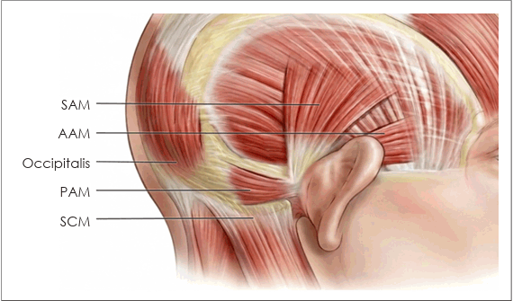
Schematic illustration of the posterior auricular muscle (PAM) and adjacent musculature in the peri-auricular region. The anterior (AAM) and superior (SAM) auricular muscles complete the extrinsic auricular muscle group, and the sternocleidomastoid muscle (SCM) and occipitalis are shown as adjacent structures.
The differential diagnosis of focal involuntary movement in the peri-auricular region includes hemifacial spasm, auricular myoclonus, and moving ear syndrome, each of which was considered and excluded in our patient. Hemifacial spasm typically involves multiple branches of the facial nerve with synchronous contractions of the orbicularis oculi, perioral muscles, and platysma [8], none of which were observed. Auricular myoclonus characteristically presents as rhythmic, often audible movements of the entire auricle accompanied by clicking tinnitus [5], whereas our patient showed irregular, non-rhythmic twitching strictly confined to the retroauricular soft tissue. Moving ear syndrome involves continuous or semi-rhythmic movement of the auricle itself driven by several auricular muscles acting in combination [4], which was not observed. The strict spatial confinement, the absence of facial or auricular co-activation, the posture-dependent triggering, and independent confirmation by three senior otologists collectively supported the clinical diagnosis of isolated postauricular muscle myokymia.
Medical therapy with anticonvulsants or benzodiazepines has shown inconsistent efficacy in auricular movement disorders [2,5]. In contrast, botulinum toxin has demonstrated favorable outcomes across conditions involving extrinsic auricular muscles, including ear-wiggling tics, muscular tinnitus, and auricular dystonia [3-6]. Reported doses vary considerably, reflecting differences in muscle involvement and localization accuracy. Because the posterior auricular muscle is extremely small—approximately 1.5-2 cm in length and 1-2 mm in thickness—limited toxin is required for effective chemodenervation [1,4]. Based on these anatomical considerations, we used a low dose (10 U total, 5 U per point) delivered as a concentrated, low-volume injection (0.1 mL; 50 U/mL). This approach was selected in an attempt to confine the toxin to the target muscle in this anatomically compact region. However, this single case does not provide comparative evidence of superiority over conventional dilution protocols, and the theoretical advantages of a concentrated, low-volume injection should be interpreted as anatomically motivated considerations only.
This low-volume approach is important because diffusion to adjacent structures, such as the sternocleidomastoid or occipitalis, may result in undesirable effects including neck weakness or scalp tightness [7]. It has been suggested that higher toxin concentrations with lower injection volumes may help limit lateral diffusion, although direct evidence remains limited and most data are extrapolated from studies on other facial and cervical muscles [4,7]. The rapid symptom resolution in our patient after a single injection session aligns with previously reported treatment responses [1,4-6]. Quantitative improvement further supports therapeutic effectiveness. No adverse events were observed, consistent with prior studies showing excellent tolerability of botulinum toxin in peri-auricular applications when dosage and injection plane are well controlled [4,7,9].
A limitation of this report is that electromyography or ultrasound guidance was not used to confirm the exact muscle of origin. However, the movement was strictly localized and clinically palpable along the posterior auricular muscle, and the robust treatment response strongly supports the accuracy of the clinical diagnosis. The pharmacologic trial of oxcarbazepine was also limited to a low starting dose without subsequent titration, although focal peri-cranial hyperexcitability syndromes such as facial myokymia have been reported to respond to lower doses than those required for seizure control [10]. Regarding long-term outcomes, complete symptom resolution was maintained at 3 months, and only minimal recurrence (VAS 1) not requiring intervention was noted at 6 months. This sustained benefit extended beyond the typical duration of botulinum toxin effect on striated muscle (approximately 3-4 months), possibly reflecting a prolonged pharmacologic effect in this small, superficial muscle. We plan to perform repeat injection sessions using the same protocol only if the patient reports a symptomatic return of the involuntary movements.
This case adds to the limited literature regarding postauricular muscle disorders and suggests that a low-dose, anatomically targeted botulinum toxin injection may be a practical and effective therapeutic option.
In conclusion, this case demonstrates that isolated postauricular muscle myokymia can be successfully managed in a single injection session through a low-dose (10 U) botulinum toxin injection. Given the small size and superficial location of the posterior auricular muscle, a concentrated, low-volume formulation was selected based on anatomical considerations. This strategy offers a safe clinical approach for managing focal peri-auricular movement disorders when the symptomatic muscle is accurately identified.
Supplementary Materials
The Supplement is available with this article at https://doi.org/10.3342/kjorl-hns.2026.00437.
Notes
Acknowledgments
This work was supported by a 2-year research grant from Pusan National University.
Author Contribution
Conceptualization: Se-Joon Oh. Funding acquisition: Se-Joon Oh. Investigation: Han-Seul Na, Se-Joon Oh. Supervision: Sung-Won Choi, Se-Joon Oh. Validation: Seokhwan Lee, Sung-Won Choi. Visualization: Han-Seul Na. Writing—original draft: Han-Seul Na. Writing—review & editing: Se-Joon Oh.